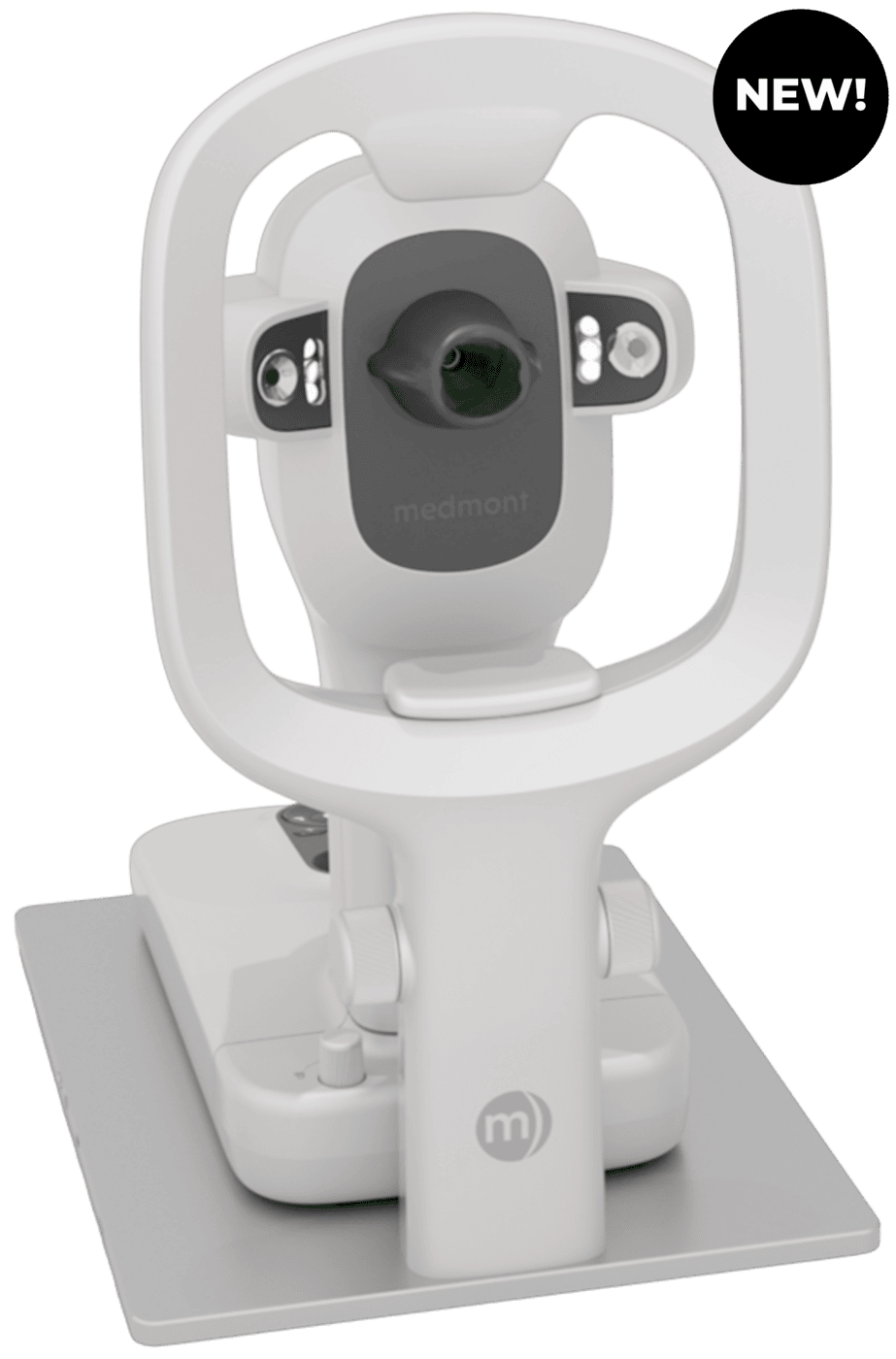
The Medmont Meridia™ Professional (Pro), distributed by NIDEK Inc., is a profitable investment. It gives you considerable value for money—over and above, in direct and indirect returns.
When looking at a corneal topographer from a profitability perspective, consider its other functions as they relate to services you can offer your patients.
To help with your research, this article explores and outlines the direct and indirect revenue you can generate with the Medmont Meridia™ Pro.
Read on to discover the Meridia Pro functions and applications designed to maximize your revenue.
What makes the Meridia™ Pro work for you?
From custom contact lenses and ortho-k fittings to corneal disease detection and ocular health monitoring, dry eye assessments and specialized treatments, the Meridia™ Pro proves its worth in every aspect of anterior eye care.
In a recent article published in Clinical Optometry, Dr Daddi Fadel refers to it as, “a state-of-the-art multipurpose diagnostic device with corneal topography and cutting-edge features.”
She writes, “[The Meridia™] consolidates a multitude of examination tools in a single instrument, enhancing practice efficiency and elevating patient care and communication. Its versatility and accuracy make it an invaluable asset in optometric practices worldwide.”
Advanced topography for custom contact lenses
Wide corneal topography coverage is essential for fitting custom lenses—and especially ortho-k. The more accurate the topography at critical lens landing zones, the higher your first-fit success rate is likely to be.
With up to 11mm of real corneal data in a single automatic capture (larger than the typical wide-cone Placido range of 6mm-9mm), the Meridia™ Pro generates an accurate baseline map, giving you real corneal data in the range that ortho-k lenses land. This is a massive advantage of the Meridia ™ Pro. It’s rare for a topographer to boast such ultra-wide capture; others leave you guessing about crucial information.
For even more data, use the Composite Mapping feature for limbus-to-limbus coverage. This extended range provides precise data for fitting, especially in the mid and peripheral regions of the cornea, as well as for scleral lens parameters.
To further aid lens parameter decisions, the Meridia™ Pro’s corneal topography data gives you accurate measurements of the horizontal visible iris diameter (HVID) and pupil diameter.
Convenient lens design options
Rounding out its position as the leading topographer for specialty lens practices, the Meridia™ Pro offers convenient lens design options. Use its built-in contact lens simulator, integrate with third party design software, or send your maps to lab consultants directly from the software.
To reduce refits and increase your chance of success, simulate corneal, ortho-k and scleral lens fits over a captured topography image, using the contact lens module. This reduces costly chair time and increases patient satisfaction.
For post-fit analysis, particularly for ortho-k, use Subtractive Maps to track corneal shape and power changes over time. You can also use them to assess corneal flattening after refractive surgery, and to monitor keratoconus advancement and locate cone position changes.
For more information on the Medmont Meridia’s contact lens features and functions, read our blog Built for first-fit success: how the Medmont Meridia™ helps you master contact lens fitting.
So, how quickly can you recoup your investment?
Looking at orthokeratology, it can cost patients $1,500 USD and more for a pair of ortho-k lenses in a treatment plan. At this rate, it would only take around 20 ortho-k patients to recoup your investment. Everything on top of this is profit.
Versatile features for more service offerings
Beyond its gold-standard limbus-to-limbus topography, the Meridia™ Pro has an HD color camera and three light sources for assessing dry eye, contact lens fits and anterior pathologies.

It’s easy to capture crisp details with the Meridia™’s HD color camera and its 2-10x digital zoom. That means you can entrust your less-costly technician with imaging, freeing you to spend quality time with your patients.
The clarity of the Meridia Pro’s images and video aids well-informed clinical decisions, clear documentation and high confidence in your treatment plans. Use them to aid patient understanding and compliance, with visual reports, allowing you to sell more treatments.
Case in point, Adrian Rossiter BScOptom, MOptom, MBA, FACBO of For Eyes Optometrists told us:
“In addition to dry eye, the Meridia™’s accuracy, large field of topography capture, ease of anterior eye and fluorescein imaging is impressive. The imaging capability is helpful for monitoring and explaining anterior eye pathology. It also makes the evaluation of RGP and orthokeratology lenses effortless.”
Additionally, you can export files to peers for a second opinion or to a specialist, such as an ophthalmologist, for collaboration and business-boosting referral opportunities.
You can also send files to contact lens labs for fast, effortless lens design or troubleshooting. In doing so, you’re supported to achieve excellent patient outcomes, ultimately increasing patient trust and positive word of mouth.
How does the Meridia Pro fit into your workflows?
The Meridia™ Pro is a versatile workhorse. It promotes good outcomes and good income. Use it throughout the spectrum of all eye exams.
Dr Aaron Wolf (OD, FAAO, FSLS, FIA, OMC) uses the Medmont Meridia™ Pro in his practice Austin Optometry Group in Austin, Texas. Dr Wolf specialises in dry eye syndrome, ocular surface diseases, and specialty contact lenses for difficult-to-fit eyes. His workflows below show how useful the Meridia™ Pro is for comprehensive, injury, dry eye and contact lens exams. It seamlessly integrates into your daily workflows, and it provides additional information to better inform diagnosis and management, making it an indispensable tool.
Comprehensive eye exam workflow:
- Infrared imaging (pupil size and meibography)
- Anterior imaging (external examination of anterior structures)

Dry eye exam workflow:
- Non-Invasive Tear Break Up Time (NIBUT) test
- Meibography
- Fluorescein imaging
- Anterior imaging (with and without Lissamine Green)

Red eye/injury exam workflow:
- Anterior imaging
- Fluorescein imaging

Specialty contact lens consult workflow:
- Infrared imaging (pupillometry and meibography)
- Anterior imaging
- Fluorescein imaging (with contact lens on eye if RGP)
- Corneal topography

Orthokeratology fitting workflow:
- Corneal topography (pre- and post-treatment)
- Fluorescein imaging (with lens on eye)

Where do Meridia™ Pro returns come from?
Besides the significant cost savings that come with lens refit reduction and reduced incorrect diagnosis, the potential for direct and indirect returns with the Meridia™ Pro is substantial.
With the Meridia™ Pro, your direct revenue comes from insurance billing and self-pay for topography and the various types of anterior imaging. Of course, insurance schemes and rebates vary depending on your country and location.
Your indirect revenue comes from specialty lens and medical care opportunities.
Thanks to the Meridia™ Pro’s crisp clear images and video, dry eye treatments such as gland expression and IPL are much easier to sell—and treatments such as drops, supplements and moisture goggles are much easier to prescribe. Watch compliance skyrocket when you use the Meridia™’Pro’s standardized visual reports as evidence.
As you can see below, there are vast indirect revenue opportunities.
| Direct returns | Indirect returns |
|---|---|
| Ophthalmological visits | Tear Osmolarity |
| Corneal topography | Immunoassay Testing |
| Anterior photography | Punctal Occlusion |
| Tear film imaging | Lacrimal Duct Irrigation |
| Meibomian gland imaging | Amniotic Membrane |
| Lid Debridement | |
| Meibomian Gland Expression | |
| IPL | |
| LLLT | |
| RF | |
| Warm Compresses | |
| Moisture Goggles | |
| Omega Fatty Acid Supplements | |
| Artificial Tears | |
| Lid Hygiene Products | |
| Eyelash Growth Serums | |
| Cosmetics | |
| Soft Contact Lenses | |
| Corneal GP Lenses | |
| Scleral Lenses | |
| Patient Referrals |
To give you an idea of your potential profit, we asked practice owner Dr Aaron Wolf OD to share his region’s care management service (CMS) fee schedule with the Medmont Meridia™ Pro. These demonstrate the rebate reimbursement opportunities that arise from using the Meridia™ Pro. These figures will differ for your region.
Reimbursement from Examination (2025 CMS Fee Schedule)
| Procedure | CPT Code | Reimbursement |
|---|---|---|
| New Comprehensive Exam | 92004 | $143 |
| Est. Comprehensive Exam | 92014 | $121 |
| Est. Intermediate Exam | 92012 | $85 |
| New 45-59min | 99204 | $163 |
| Est 20-29min | 99213 | $89 |
| Est 30-39min | 99214 | $125 |
Reimbursement from Imaging
| Procedure | CPT Code | Reimbursement |
|---|---|---|
| Corneal topography | 92025 | $35 |
| Anterior photography | 92285 | $22 |
Reimbursement from Non-Covered Diagnostics
| Procedure | CPT Code | Reimbursement |
|---|---|---|
| Punctal Plugs | 68761 | $139 |
| Lacrimal Duct Irrigation | 68840 | $128 |
| Amniotic Membrane | 65778 | $1,218 |
| Eyelash Epilation | 67820 | $18 |
The ‘Pro’ is for ‘profitable’!
Thanks to advanced features that enhance your diagnostic capabilities, increase efficiency and open numerous revenue streams, the Medmont Meridia™ Pro is a fast pathway to practice prosperity. And it future-proofs your business.
Moreover, investing in the Meridia™ Pro reduces the expense of lens refits and incorrect diagnosis. According to Dr Daddi Fadel, “Having such a plethora of examination tools in one instrument increases practice efficiency and enhances the care of patients, their education, and compliance with clinician’s recommendations. Furthermore, this integration not only saves chair time and space but also proves to be a more economical investment”.
RELATED VIDEOS
Video transcript
Background music plays throughout the video. There is no spoken narration — the video consists of on-screen text and product visuals of the Medmont Meridia corneal topographer.
Text: "Over Twenty Years Building a Legacy in Topography Precision..."
Text: "...Now Continues with... More Clarity in your approach"
Text: "More Convenience in your workflow, More Confidence in your decisions"
Text: "One System. Many Choices."
The Medmont Meridia device is shown from the front, with the text "medmont meridia™ Advanced Topographer."
The device is shown at a closer angle, labeled "medmont meridia™ | Professional."
The device is shown again with the text "See Clearly, Treat Optimally."
A close-up of the device's scanning head is shown, alongside a color corneal topography scan image (a rainbow-colored map of the cornea). Text: "Color Topography Placido & Profile."
The scanning head is shown next to a close-up photo of a human eye. Text: "Anterior."
The scanning head is shown next to an eye image with a green fluorescein dye pattern. Text: "Fluorescein."
The scanning head is shown next to a black-and-white image of an eyelid. Text: "Meibomian."
The scanning head is shown next to a black-and-white eye image with two light reflection points measured. Text: "Tear Meniscus Height."
A close-up shows a calibration compartment on the device opening to reveal an internal calibration piece, with a separate cover panel beside it. Text: "Sensible, Secure, and Simple Calibration Storage."
A close-up of the device's control panel shows five illuminated navigation buttons (arrows and shape icons). Text: "Ergonomic Navigation with Illuminated Quick Keys."
Text: "Clarity Simplified - Medmont Studio 7," with a blurred monitor graphic in the background.
A monitor displays the Medmont Studio 7 software interface, showing a patient list and an "Exam Type" selection screen with icons for Topography, Composite Topography, Video Topography, Tear Film Analysis, Anterior Capture, Fluorescein Capture, and Meibomian Gland. Text: "Clarity Simplified - Medmont Studio 7."
Two sample printed patient report pages are shown: a "Medmont Meridia Dry Eye Report" with patient information, diagnostic scores, and eye images, and a second page with additional eye photos (limbal redness, blepharitis), test results, and a summary with diagnosis and treatment recommendations. Text: "Engaging Patient Dry Eye Reports."
The full device is shown again with the text "medmont meridia™ Advanced Topographer," along with a regulatory caution notice at the bottom: "Caution: U.S. Federal Law restricts this device to sale, distribution, and use by or on the order of a physician or other licensed eye care practitioner. Specifications may vary depending on circumstances in each country. Specifications and design are subject to change without notice." Dated November 13, 2020, document number 2020-0013.
The device is shown with the text "Now Available," along with the same regulatory caution notice and date.
The device is shown with the text "Know more. Know now. www.medmont.com," along with the same regulatory caution notice and date.
Video transcript
Welcome to this introduction to the Medmont Meridia Advanced Topographer. This exciting instrument allows you to perform corneal topography and multiple imaging options that make for an incredibly powerful instrument in your practice today.
The Medmont Meridia is many instruments in one. It's first and foremost a corneal topographer, but the imaging options allow it to take anterior segment white light captures, non-invasive tear film breakup time testing, meibography, as well as fluorescein assessment — with the patient behind this one single instrument, there's so many things you can do.
The Medmont's cone construction and focal distance allow for incredibly large surface areas of capture. The vast majority of the cornea can be taken with one single topography, and that gives you a very complete assessment of the corneal shape, power, and elevation of the eye. But the unique Medmont composite eye feature allows you to expand an already large surface area of capture to create a complete view of the corneal surface from limbus to limbus, and this can be beneficial in the fitting of specialty contact lenses that may be as large as the corneal diameter or larger.
The Medmont includes multiple disease detection indices that will assist you in diagnosing cases that may require closer follow-up, treatment, or surgery. The contact lens software is one of those unique features of the Medmont that allows you to build your specialty contact lenses from the topographies. You can build your corneal GP single vision and multifocal lenses, your ortho-K, your scleral, and many other specialty lenses for your patients. In orthokeratology and myopia control analysis, the Medmont's subtractive maps can be an important tool in your diagnosis of the outcomes following lens wear.
The Medmont Meridia offers an entire dry eye assessment suite to assist you with your diagnosis, your treatment, and your ongoing care of your dry eye patients. Placido technology will allow us to reflect the rings off the tear film to have an appreciation of the tear film quality — this also allows us to have an appreciation of the non-invasive tear film breakup time for each eye. The assessments that the Medmont Meridia will do to assist with your dry eye analysis are fully automated and something that's quickly and efficiently performed on the eye.
The Medmont Meridia will also allow for an assessment of the tear meniscus height, again to assist with your overall dry eye assessment for this patient. And another very valuable dry eye assessment is the infrared meibography that your Medmont Meridia can perform.
The instrument is also able to take anterior segment white light imaging — this can be an important way to document findings from each of your visits. This white light imaging can be helpful in your contact lens practice as a way that you can share the information with your lens consultant to best optimize the fit of your lenses. Similarly, your Medmont Meridia is able to perform a fluorescein assessment of the cornea or of your rigid lenses.
With all of the imaging options in your Medmont Meridia, you have the ability to take single images or videos in any of the capture modes, be it the fluorescein, the meibography, or the white light capture. With this one powerful instrument, you have all of your images, videos, and captures available in one place, in one software, making it simple and efficient to review and assess.
Additionally, the Medmont Meridia makes it possible for you to create a comprehensive dry eye report. You have the ability to create a custom template specific for your practice and your areas of analysis and assessment. This dry eye report can become an important communication tool that you use with your patients to assist in their understanding of both the diagnosis as well as the treatment plan. And with any of the captures that you take with your Medmont Meridia, you're able to quickly and efficiently export the file so that it can be emailed or sent to your specialty lens consultant when you need assistance with a particular case.
The Medmont Meridia has always been known as the ultimate specialty contact lens tool, one that provides a large surface area of capture and accurate information so that we can best design our initial specialty contact lenses. But we also now have the ability to add an entire dry eye assessment to our practice. Additionally, we have numerous ways that we can now image and record video of the anterior segment in white light, in infrared, or in fluorescein.
The Medmont Meridia is so much more than just a corneal topographer — it's also an incredible imaging device that can be used for so many functions and applications in your practice. With the Medmont Meridia, know more, know now.
For more information, go to the Medmont website at medmont.com. Thank you for your interest in the Medmont Meridia Advanced Topographer.
To learn more about the Meridia topographer, please visit: https://usa.nidek.com/meridia