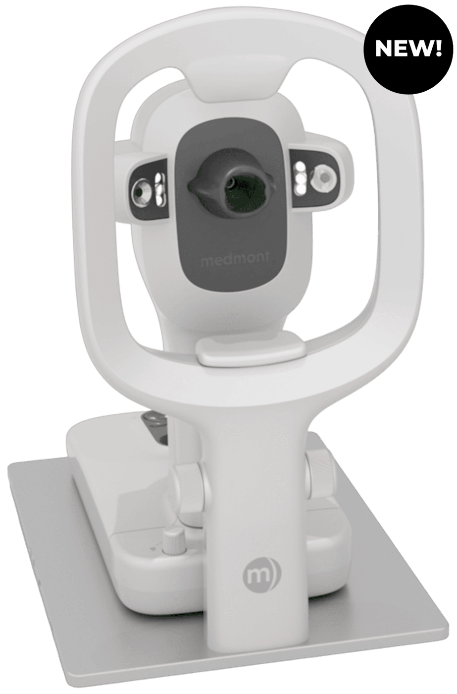
Meridia™ Vantage Overview
The Vantage Advantage

All-in-one Diagnostics
Gold-standard topography, HD colour anterior imaging, and dry eye analysis.
Corneal-scleral Topography
Capture up to 20mm × 18.5mm in a single capture — no stitching. High fit accuracy, fewer remakes, complete data for large-diameter lenses.
Fast, Intuitive Workflow
Instant capture, rapid analysis, single-operator design and patient-friendly ergonomics.

Enhanced Applications
Full color images for corneal topography, specialty lens fitting, anterior and fluorescein images, and dry eye evaluations.
Corneal Coverage
Using 32 rings with 102,000 measurement points, the meridia provides detailed topography data over a wide area of the human cornea.
User Friendly
Ergonomic design with 5 quick keys are designed to improve workflow efficiencies and user experience for you and your staff.
Meridia™ Classic + Professional Overview
First in Class Accuracy
Medmont Meridia: Advanced Corneal + Scleral Topography Platforms
| Key Features |  Classic |
Pro |
 Vantage |
|---|---|---|---|
| Increased Field of View Topography | Yes | Yes | Yes |
| Optimized Depth of Field Focus | Yes | Yes | Yes |
| Miniaturized AC/DC Adapter | Yes | Yes | Yes |
| 3.0/3.1 USB Interface | Yes | Yes | Yes |
| Ergonomic Quick Keys | Yes | Yes | Yes |
| Convenient and Secure Calibration Ball Storage | Yes | Yes | Yes |
| High Resolution Digital Color Imaging | Yes | Yes | Yes |
| Automated Horizontal Visible Iris Detection | Yes | Yes | Yes |
| Scleral Lens Fitting Simulation | Yes | Yes | Yes |
| Improved User Experience and Interface with Studio Software v7 | Yes | Yes | Yes |
| Anterior Imaging and Video | No | Yes | Yes |
| Meibomian Gland Imaging | No | Yes | Yes |
| Fluorescein Imaging and Video | No | Yes | Yes |
| Tear Meniscus Measurements | No | Yes | Yes |
| Corneal Staining and Fluid Dynamics | No | Yes | Yes |
| Scotopic and Photopic Pupil Measurements | No | Yes | Yes |
| Focus Guidance Aid | No | Yes | Yes |
| Imaging Grading Scales | No | Yes | Yes |
| Screening Reports | No | Yes | Yes |
| Scleral Topography | No | No | Yes |
Experience Gold Standard Topography
Advanced Corneal + Scleral Topographers
The Medmont Meridia™ Vantage has become an essential part of my clinical workflow. I primarily use it for scleral lens fitting, orthokeratology, and dry eye evaluations.
It has reduced the number of trial lenses I need for sclerals, allows me to clearly monitor ortho-k treatment patterns, and provides objective imaging for dry eye that makes patient education much easier.
It’s reliable, intuitive to use, and has genuinely elevated the level of care I’m able to provide in my practice.

Hanish Patel OD, FAAO
Eye Associates Of New York/Center for Ophthalmic and Vision Research
Get Started
Take Patient Care to the Next Level
Accelerate the discovery and development of patient treatment and operate more efficiently with NIDEK’s ophthalmic solutions.