Skip to content

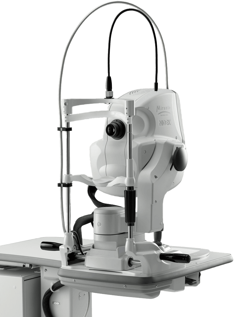
NIDEK Scanning Laser Ophthalmoscope / OCT Mirante

Overview

Nidek Scanning Laser Ophthalmoscope Mirante More details

RGB Triple Detectors

Three separate RGB detectors simultaneously scan different depths of retina with red, green, and blue wavelengths. A color histogram is available for fine adjustment based on pathology or practitioner preference.

More details

Ultra wide field x ultra HD image*

The clear image of the entire 163° field of view** enables detailed evaluation of pathologies from the fovea to the extreme periphery.

* Ultra wide field imaging is available with the optional wide-field adapter.

** Measured from the center of the eye.

More details

Retro Mode

Retro mode is a unique non-invasive technique for detecting pathologic changes in the choroid. This imaging modality uses scattered IR light to detect abnormal reflection in the choroid caused by drusen, edema and other subtle chorioretinal pathologies.

More details

Easy Tilt and Swing

The tilt and swing functions for the optical head allows imaging of the peripheral fundus and acquisition of panorama images. These functions also help for patients with unstable fixation.

Mirante Overview

State-of-the-art SLO/OCT Combo

Unsurpassed Color and Clarity for Every Detail

The clear image of the entire 163° field of view enables detailed evaluation of pathologies from the fovea to the extreme periphery. Standard field is 89°.

(Ultra wide field imaging is available with the included wide-field adapter.)

Effective Evaluation of Dynamic and Static Blood Flow

The Mirante allows simple, simultaneous acquisition of FA and ICG images.

The live IR monitoring enables alignment prior to fluorescence emission and reduces the risk of missing the very early phase of angiography.

HD 16.5 x 12 mm Wide Area Imaging with a Wealth of Functions

The maximum 16.5 x 12 mm area scan available with the Mirante allows wide area diagnosis including the macula and optic disc in a single shot. The ultra fine mode and tracing HD plus functions provide high quality images for detailed observation from the vitreous layers to the choroid.

Intuitive Functionality for Efficient Workflow

The Mirante has multiple modalities and functions with interface software that presents these choices in a simple, easy-to-use manner. This functionality streamlines the clinical workflow while capturing images with the required settings.

Promotional Poster For The Nidek Image Of The Year Award 2020. It Features Three Retinal Images, Each Showcasing Intricate Details Of The Eye. The Text Reads, &Quot;Nidek Image Of The Year 2020&Quot; And &Quot;Check Out All Submissions.
Image Award 2020
A Promotional Image For The 2Nd Annual Nidek Image Of The Year. It Features A Badge Showing &Quot;Image Award 2021&Quot; And Four Detailed Retinal Images. Below The Images, Text Reads, &Quot;Check Out All Submissions.&Quot; The Background Is Light With Prominent Gold Text.
Image Award 2021
Image Featuring The Announcement For The 3Rd Annual Nidek Images Of The Year With An &Quot;Image Award 2022&Quot; Badge. The Banner Includes Retinal Images, One In Color And One In Black And White. A Blue Text Box At The Bottom Reads &Quot;Check Out All Nidek Submissions.
Image Award 2022
Banner For The 4Th Annual Nidek Image Of The Year Contest. Text Includes &Quot;Image Award 2023,&Quot; &Quot;Mirante,&Quot; &Quot;Check Out All Submissions,&Quot; And &Quot;Nidek.&Quot; There Are Five Retinal Images Showcased. Nidek Logo Is At The Bottom Right. Background Has A Blue And White Color Scheme.
Image Award 2023

Versatile Multimodal Imaging Platform

Scanning Laser Ophthalmoscope

The Mirante platform is a versatile multimodal imaging tool which has proved to be invaluable for diagnosis, staging and management of various retinal diseases in our clinical practice. Overall, the Mirante is a powerhouse device that allows us to comprehensively assess retinal diseases in our patients.

A Person With Short, Dark Curly Hair And Glasses, Dressed In A Black Suit With A White Shirt And Red Tie, Is Smiling And Looking At The Camera. The Background, Softly Blurred With Warm Lighting, Subtly Features The Logo Of Nidek.

SriniVas R. Sadda, MD

Professor of Ophthalmology

I have been using the Nidek Mirante since 2020, and it has ever since become an essential element in my practice. The ability to perform multiple imaging modalities in a single device greatly facilitates patient assessment and optimizes my workflow, with the added benefits of unique features such as retro mode imaging.

A Man With Dark Hair And A Beard Is Wearing A Dark Suit, White Shirt, And Striped Tie. He Is Smiling Slightly And Facing The Camera Against A Plain White Background.

José Gerardo García Aguirre, MD

Ophthalmologist

Essential Element in the Practice

Scanning Laser Ophthalmoscope

RELATED PRODUCTS:

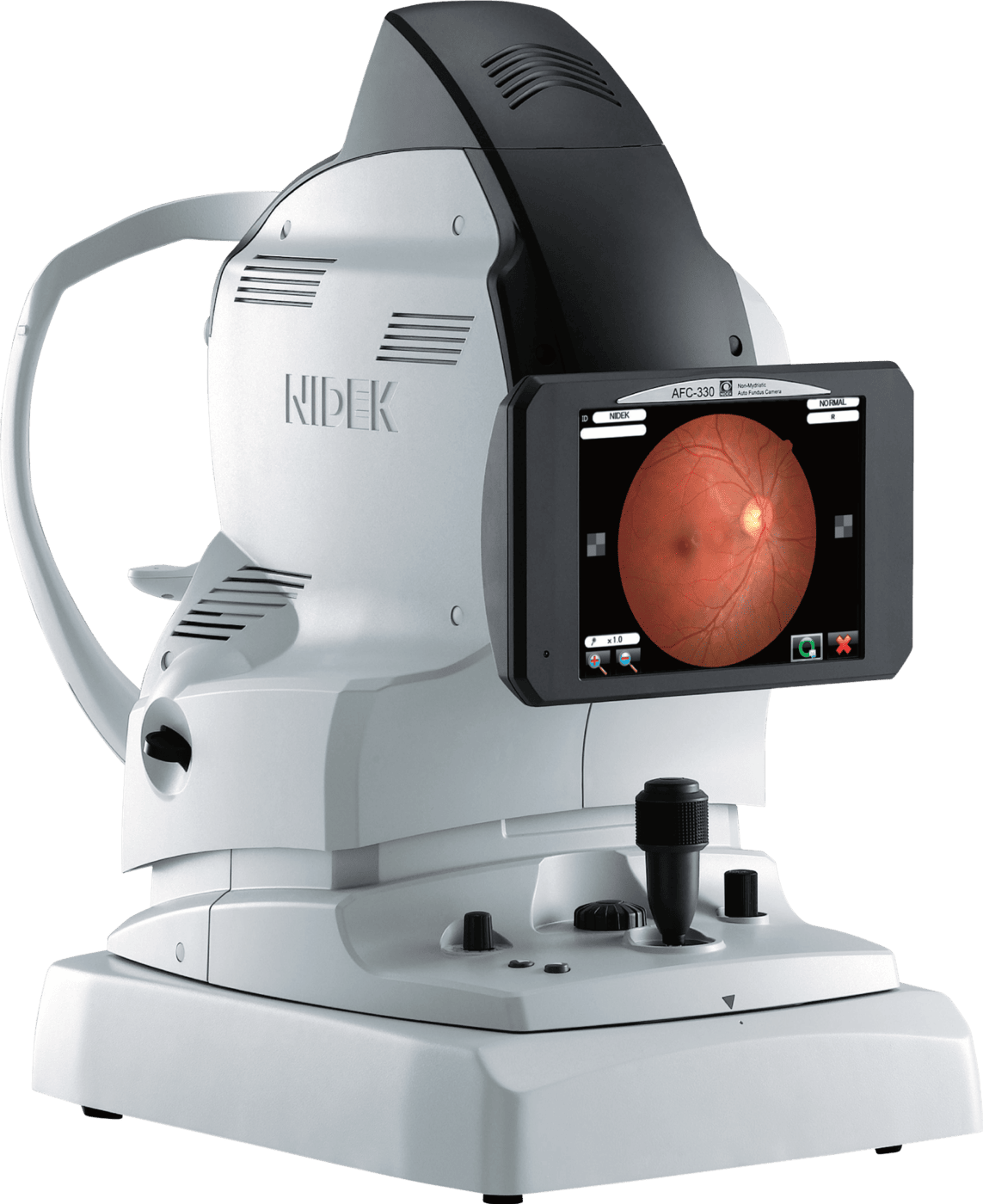
A Medical Imaging Device Used For Retinal Scanning, The &Quot;Nidek&Quot; Features A White Body With A Black Cap And A Display Screen Showing An Image Of An Eye'S Retina. It Boasts Various Controls And Adjustment Knobs On Its Base For Precision.
AFC-330 Non-Mydriatic Auto Fundus Camera
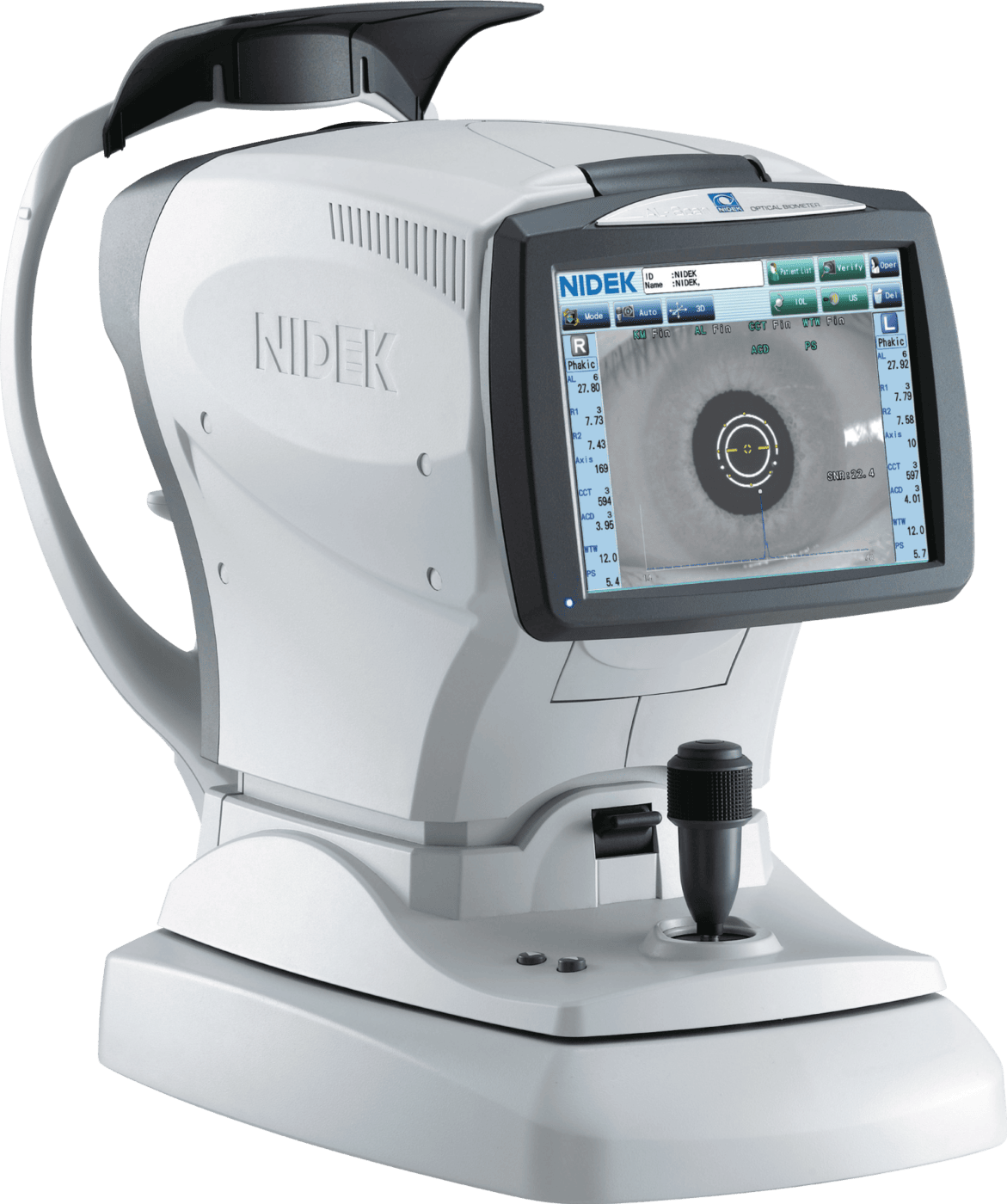
A Nidek Medical Device With A Digital Screen, Used For Eye Examination. The Machine Features Buttons, Knobs, And A Viewing Lens. The Screen Displays An Image Of An Eye, Illustrating Its Use In Ophthalmology For Detailed Eye Analysis Or Diagnostics.
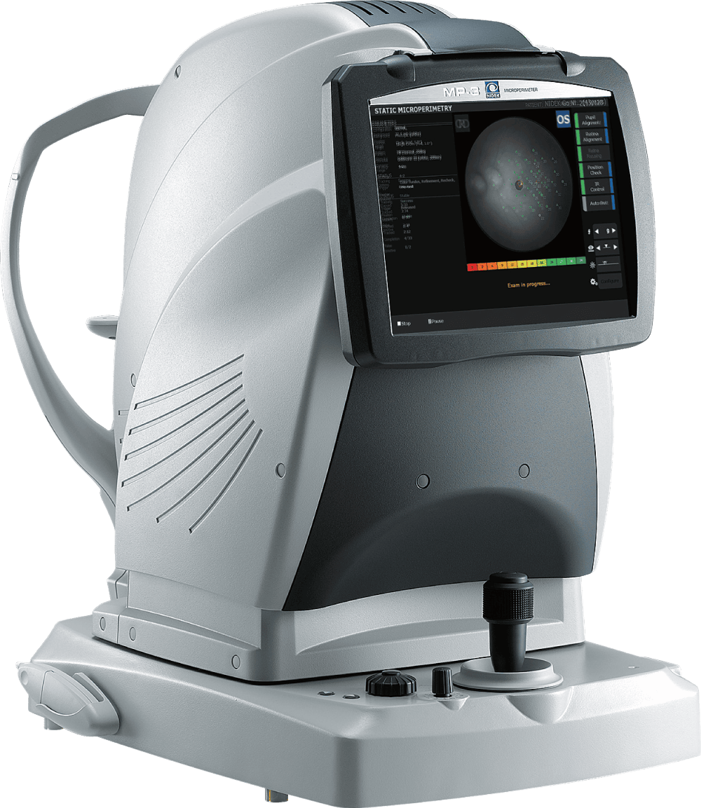
MP-3 Microperimeter
A Modern Nidek Ophthalmic Slit Lamp Biomicroscope On A Wheeled Stand With Various Attached Components, Including An Adjustable Binocular Microscope, Control Knobs, And An Integrated Light Source. A Foot Pedal Is Placed Nearby On The Floor.
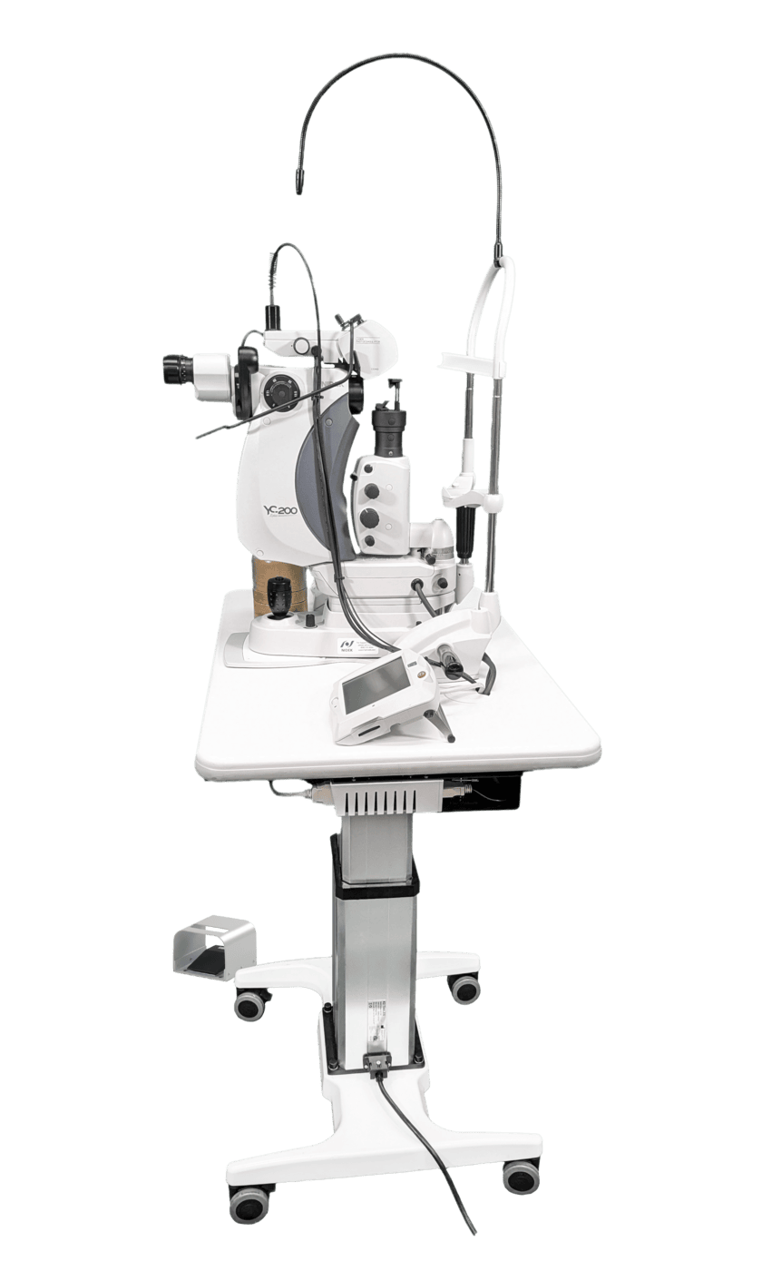
YLC-500/500 Vixi | Yellow: Laser Photocoagulator/Scan Laser Photocoagulator
An Ophthalmic Slit Lamp Biomicroscope By Nidek Is Setup On A Height-Adjustable Table, Equipped With Binocular Eyepieces, Control Knobs, And An Attached Chin Rest For Patient Use. The Table Has A Control Panel And Sits On Wheels For Easy Mobility.
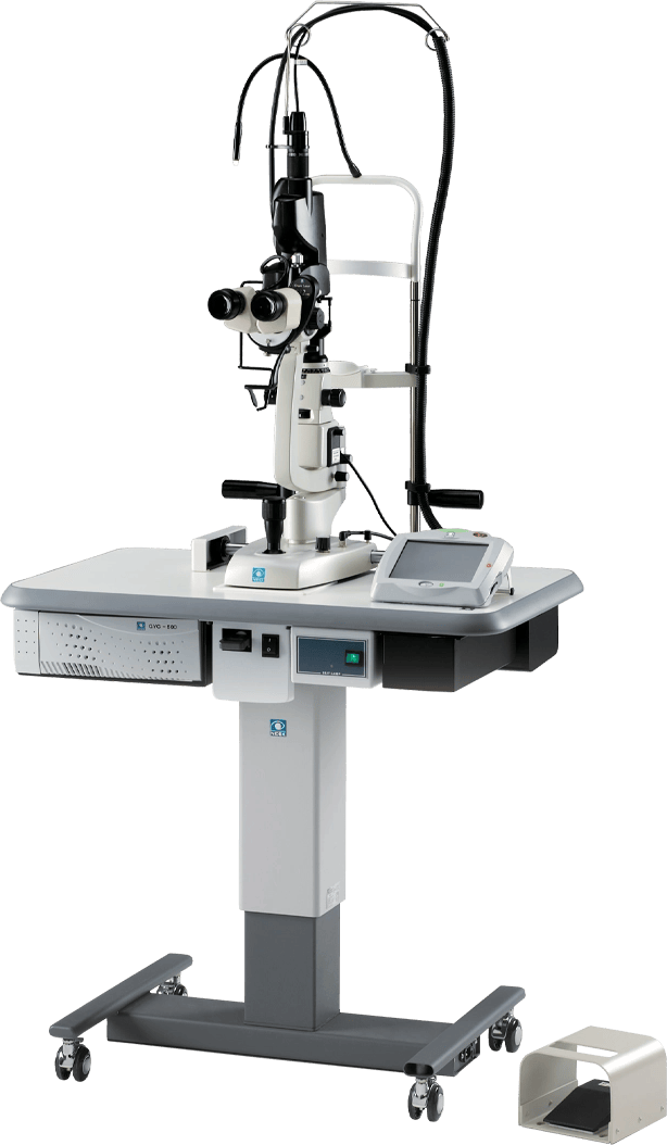
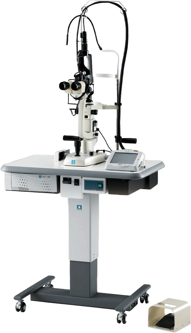
GYC-500/500 Vixi | Green: Laser Photocoagulator/Scan Laser Photocoagulator
A Nidek Slit Lamp Biomicroscope With An Integrated Computer Screen And Control Panel On A Stand. The Unit Connects To A Larger, Wheeled Device Featuring Vents And A Cylindrical Component, Possibly For Additional Diagnostic Or Therapeutic Functions.
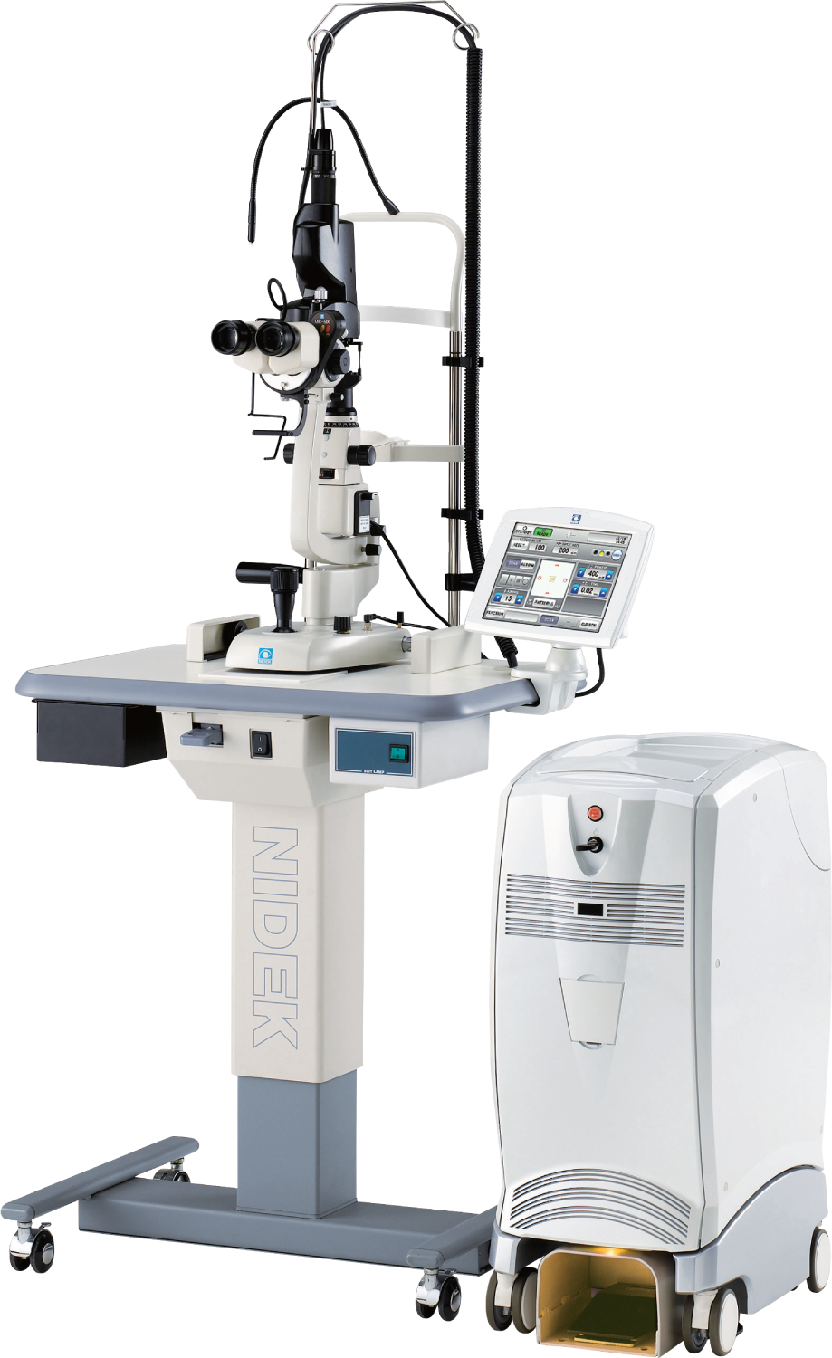
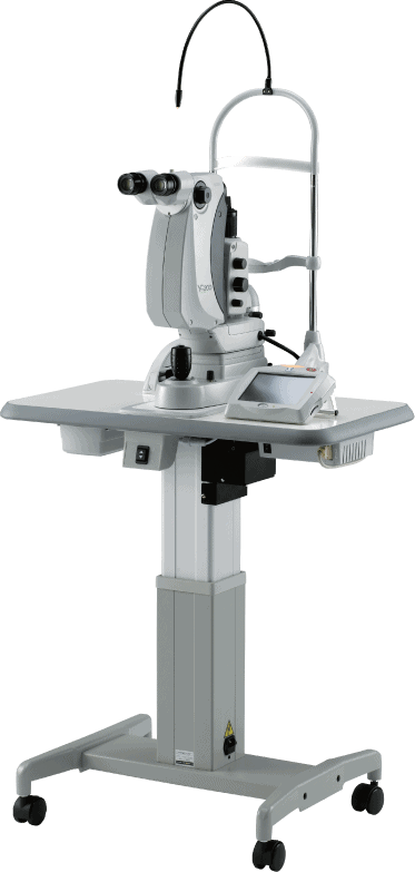
MC-500/500 Vixi | Multicolor Laser Photocoagulator: Green | Yellow | Red
NIDEK USA

Privacy Overview

This website uses cookies so that we can provide you with the best user experience possible. Cookie information is stored in your browser and performs functions such as recognizing you when you return to our website and helping our team to understand which sections of the website you find most interesting and useful.

Please visit our privacy policy for more information regarding the personally identifiable information we collect and how it is used.