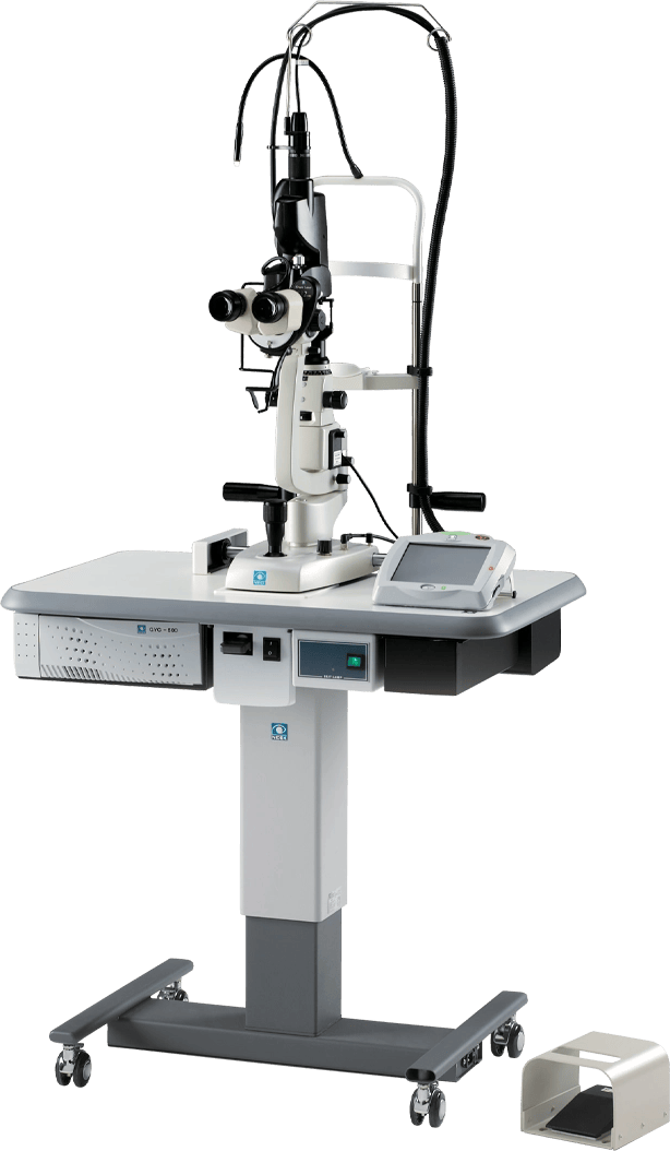
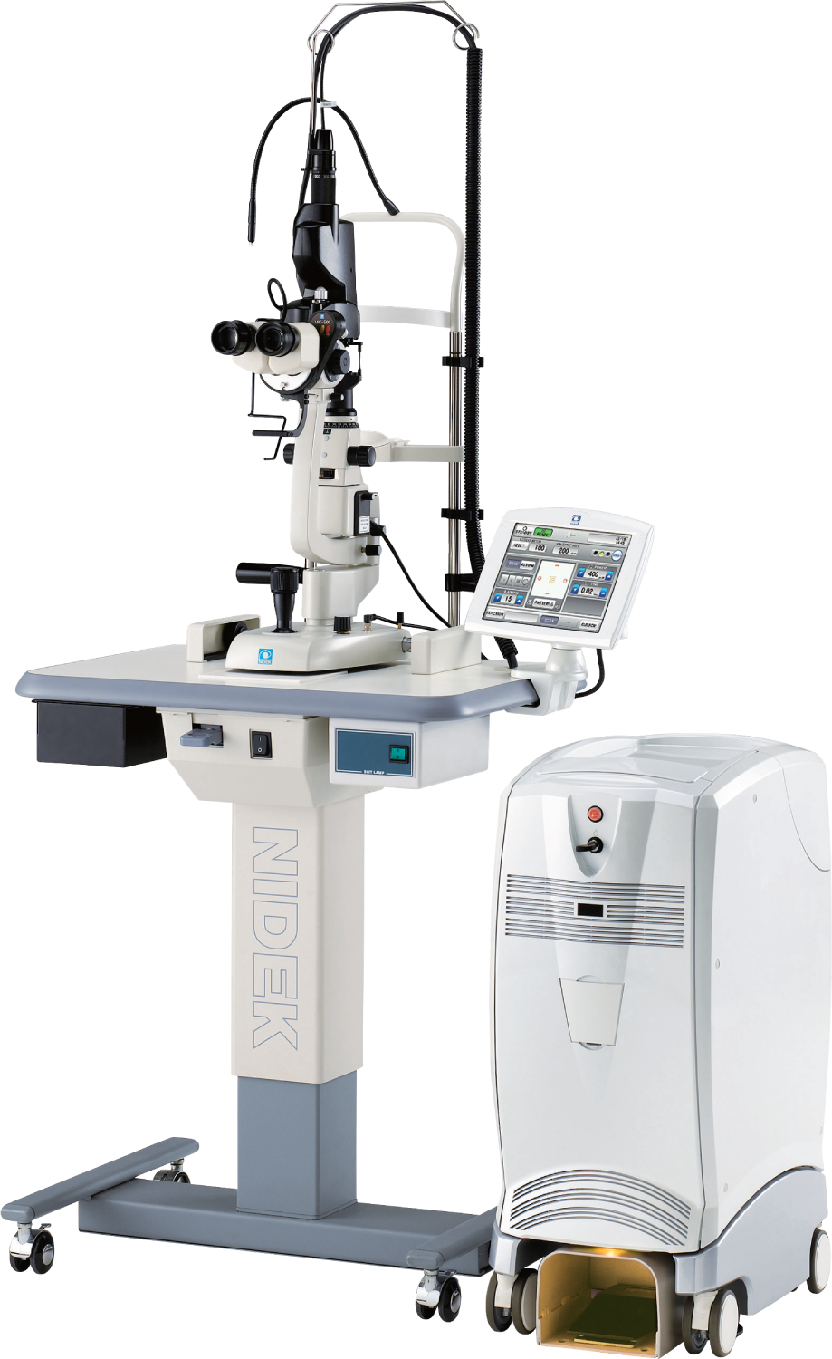
 More details
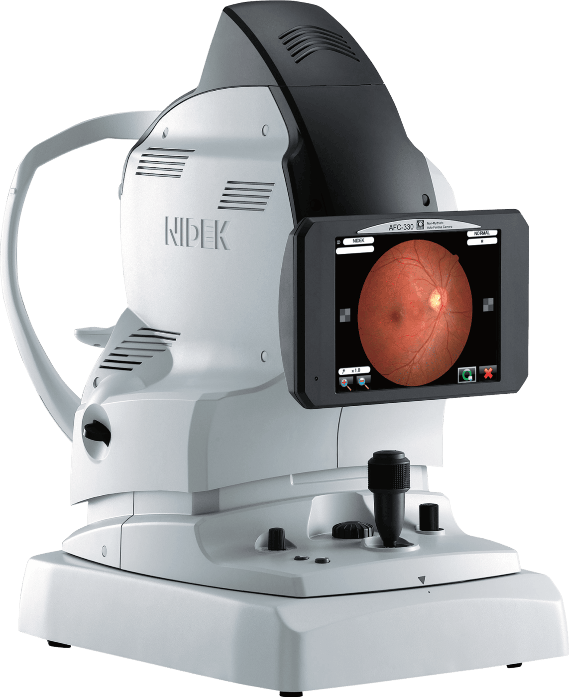
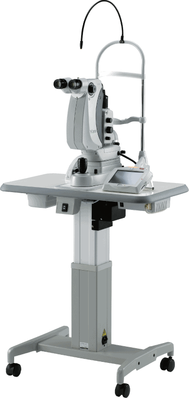
High Resolution Non-Mydriatic Fundus Camera
The 12-megapixel fundus camera in the MP-3 acquires high resolution images of retinal pathology and allows easy image acquisition
Patient Comfort
Up/Down buttons are used to adjust the motorized chin rest to the correct height for patient measurement.
Built-In Touch Screen
Large Tiltable LCD screen for superior viewing angle and operational position options.
User-Friendly Design
NIDEK’s much acclaimed 3-D auto tracking and auto shot provides the operator with the most ease, comfort, and accuracy on all measurements.
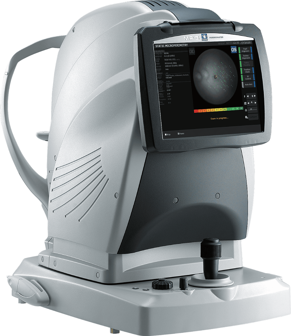
MP-3/3 type S Overview
New Paradigm in Low Vision
Trusted By Top Care Eye Professionals
Advanced Microperimeter
The NIDEK MP-3 Microperimeter has been a valuable tool in showing the patient’s functional correlation to their macular condition. I will frequently perform microperimetry for patients with dry age-related macular degeneration, macular pucker, central serous retinopathy, and other macular conditions to assess whether the anatomic findings observed on exam and OCT are causing a functional deficit.

Sadiq Syed, MD
Maryland Retina
Get Started
Take Patient Care to the Next Level
Accelerate the discovery and development of patient treatment and operate more efficiently with NIDEK’s ophthalmic solutions.