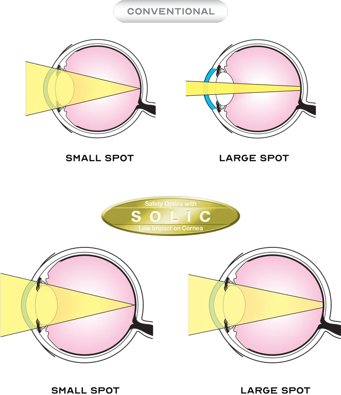
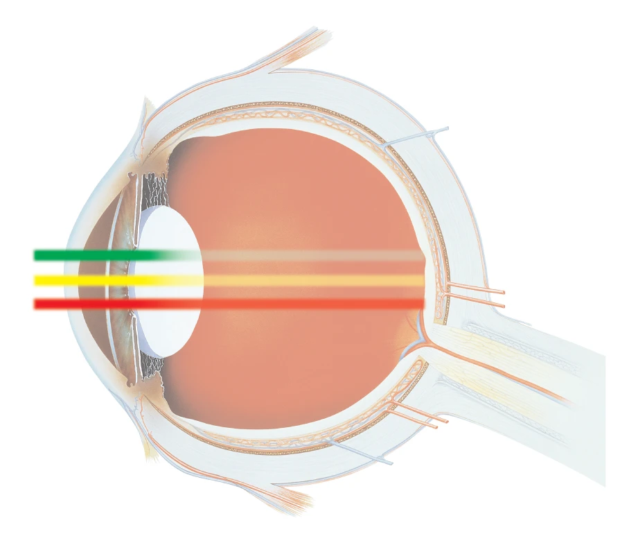
Safety Optics with Low Impact on Cornea
All delivery units incorporate the SOLIC optical design that ensures low energy density on the cornea and lens even for large spot sizes.
Multicolor Laser for Multiple Applications
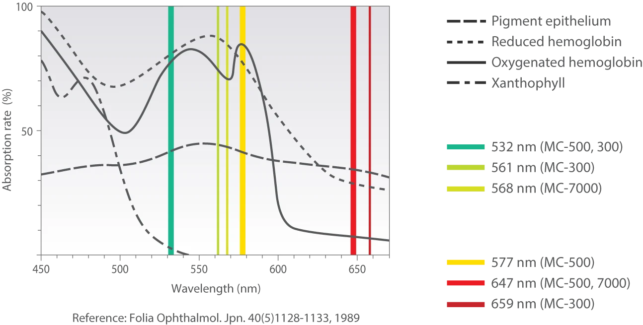
The MC-500 Vixi / MC-500 enables efficient photocoagulation even through opaque media.
In cases of cataract, better penetration is achieved with the yellow wavelength (577 nm) compared to green. In eyes with retinal hemorrhage, better penetration is achieved with the red (647 nm) wavelength.
NOW AVAILABLE WITH ALL NIDEK PHOTOCOAGULATORS
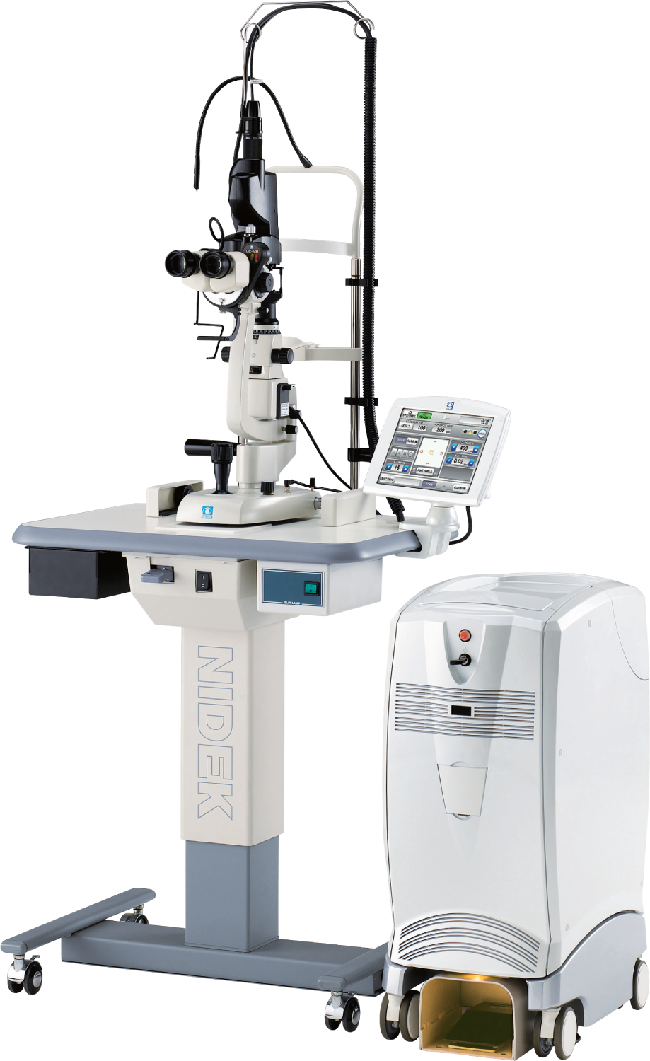
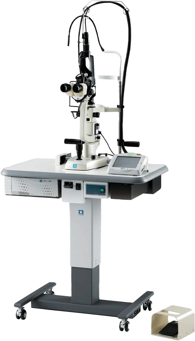
Reduce Patient Chair Time with MC-500 Vixi
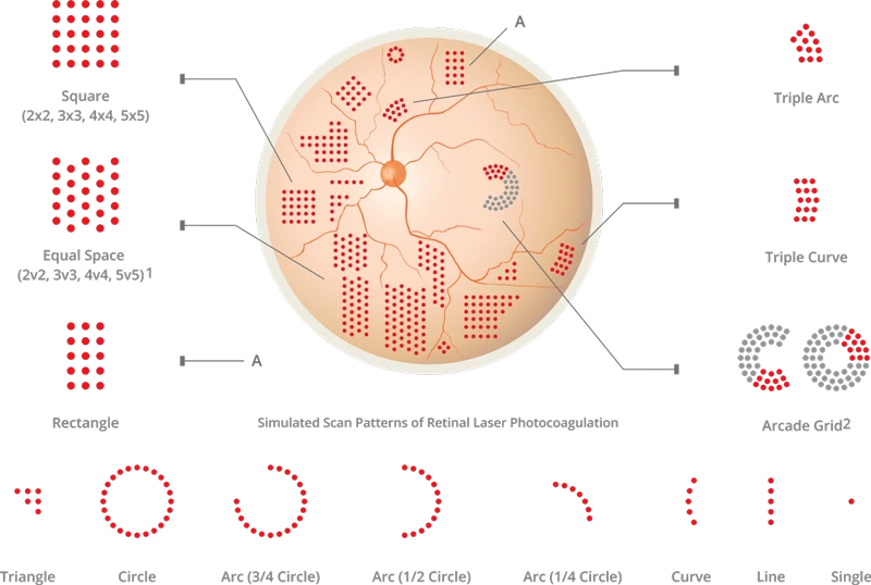
Scan delivery units with Vixi enhance treatment by its ability to treat varying retinal pathologies with 22 preprogrammed scan patterns.
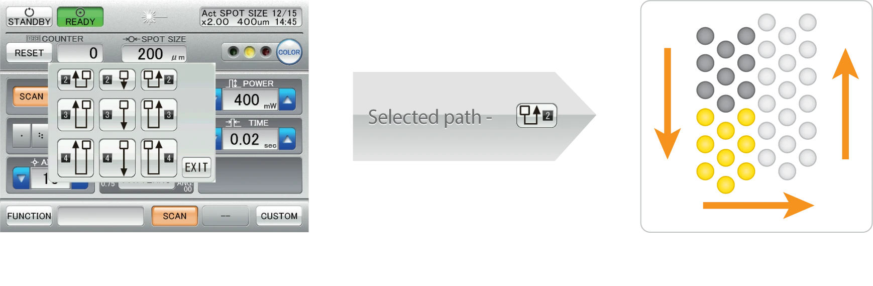
Once photocoagulation is completed in one region, the auto forward function automatically positions the scan pattern to the next region of treatment, allowing the surgeon to concentrate better on adjusting focus.
01
Spacing Function
2v2, 3v3, 4v4, 5v5 – Equal spaces between spots in all direction.
02
Auto Forward
Automated positioning of the scan pattern for photocoagulation.
03
Repeat Mode
Regions undergo photocoagulation continuously on a pre-programmed path.
Wide Range of Delivery Unit Options
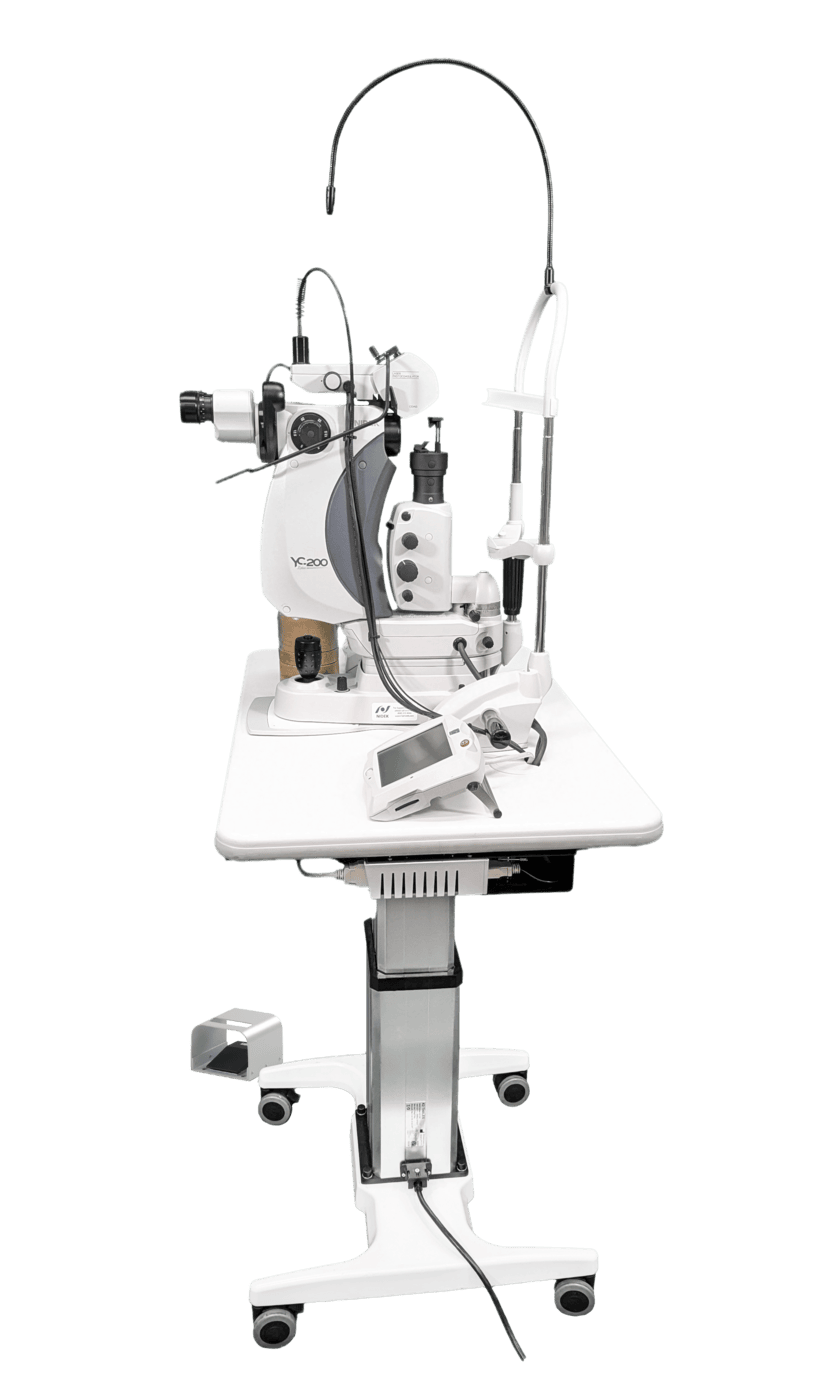
In addition to conventional single delivery units, the scan delivery units are added to the wide range of multicolor laser delivery systems. Both the scan and single delivery units include attachable models³ for the NIDEK SL-1800, ZEISS SL130, and HAAG BQ900, which provide the existing slit lamps with a new stage for scan and single laser treatment.
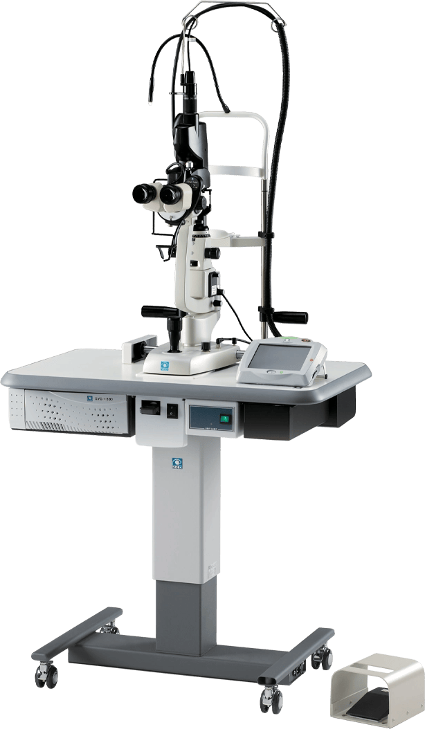
MC-500 &
MC-500 VIXI

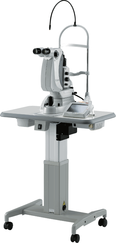
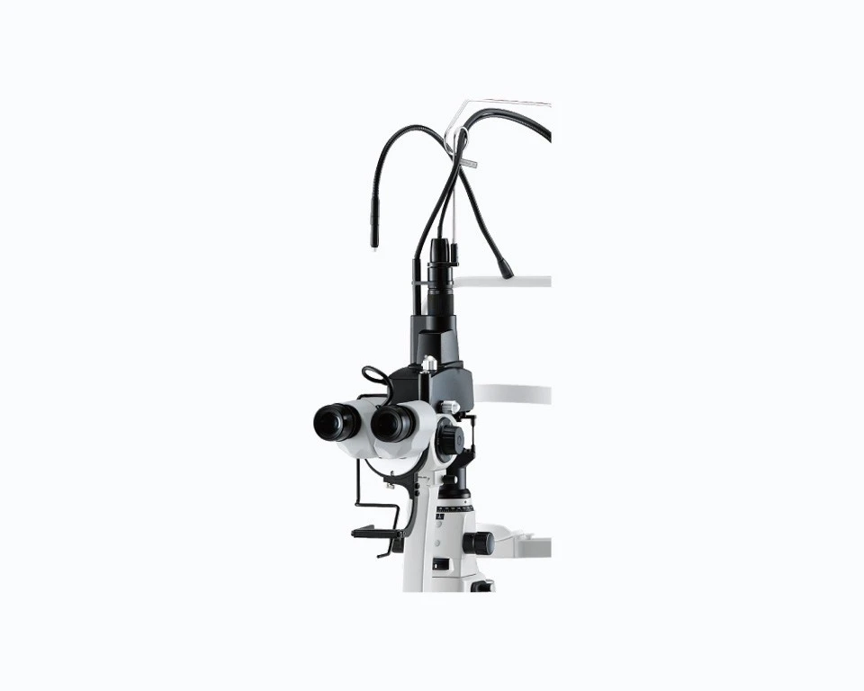
Slit Lamp Delivery Unit

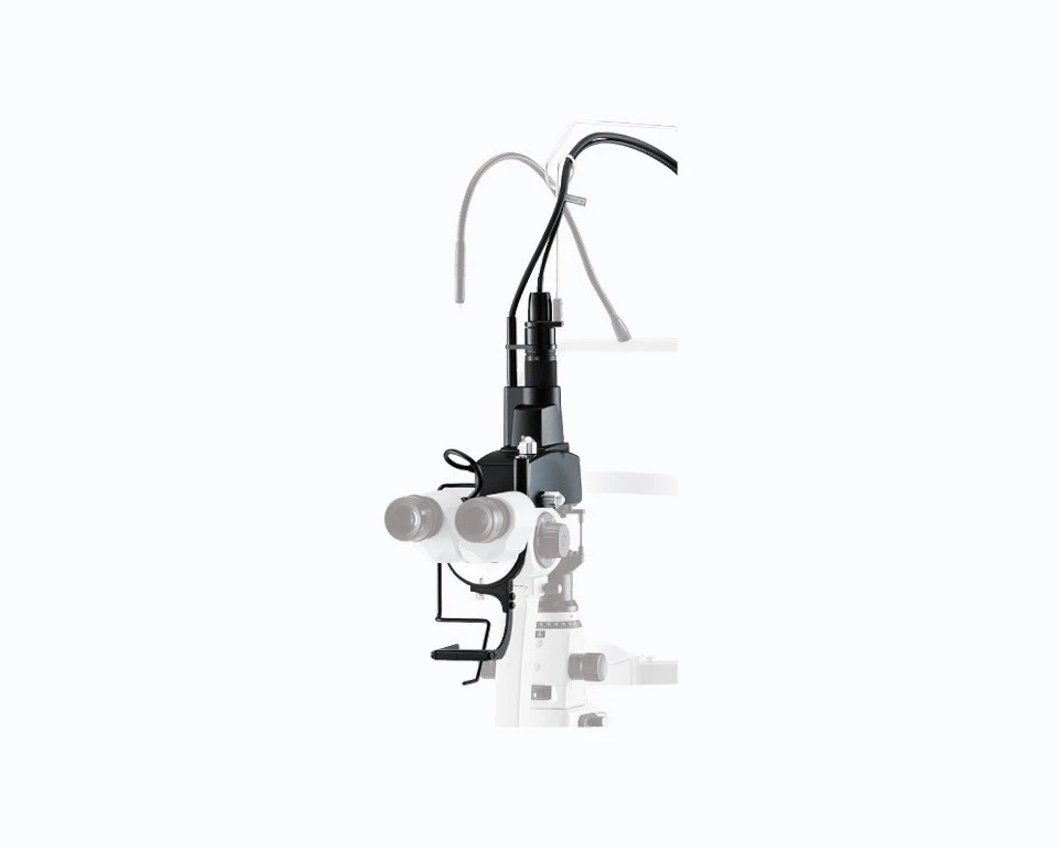
Attachable Delivery Unit
Scan Slit Lamp Delivery Unit
Scan Attachable Delivery Unit

Scan Attachable Delivery Unit

BIO Delivery Unit
Attachable Delivery Unit
Scan Slit Lamp Delivery Unit
Scan Attachable Delivery Unit
Scan Attachable Delivery Unit
BIO Delivery Unit
Slit Lamp Delivery Unit
Experience powerful multicolor laser technology and get access to premium support and resources.
Get in touch with a sales professional.
¹ For equal space patterns, No. v No. indicates the number of spots in horizontal and vertical directions.
² The arcade grid pattern is used for treatment of the periphery of macula in one-sixth units. The inner diameter is fixed and spot sizes range from 100 to 200 μm.
³ Prior confirmation of attachment to an existing slit lamp model is required.