Skip to content

NIDEK Specular Microscope CEM-530

Overview

Nidek Specular Microscope Cem-530 More details

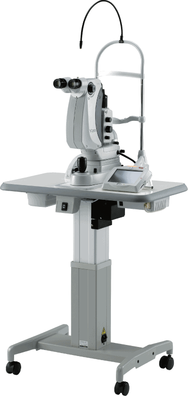
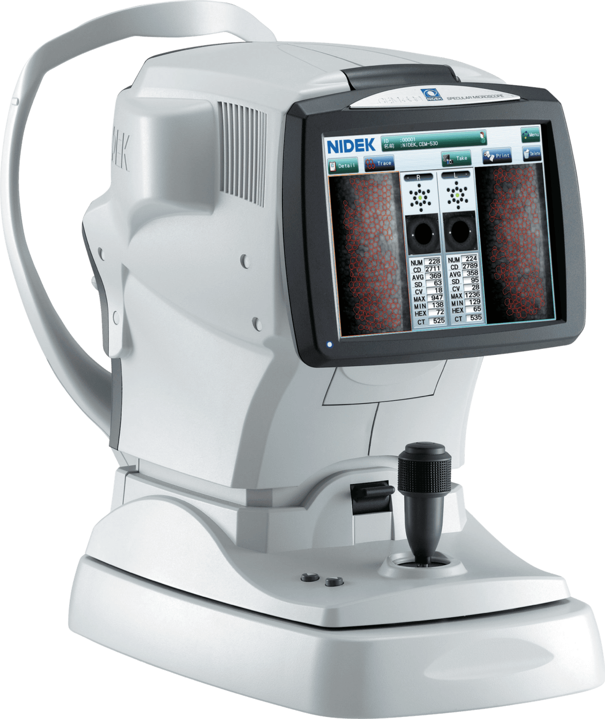
Multi Area Specular Microscopy

The combination of central, paracentral, and peripheral imaging is available.

More details

Patient Comfort

The 3-D auto tracking and auto shot, provides ease of use, allowing faster and more accurate measurement.

More details

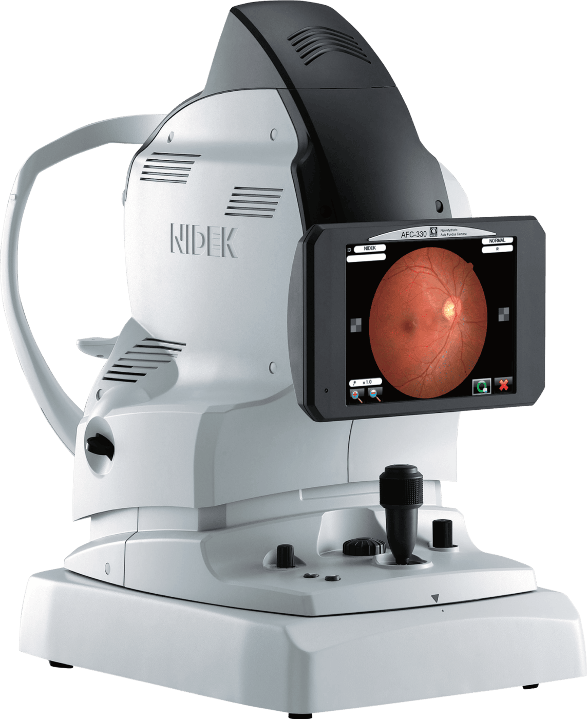
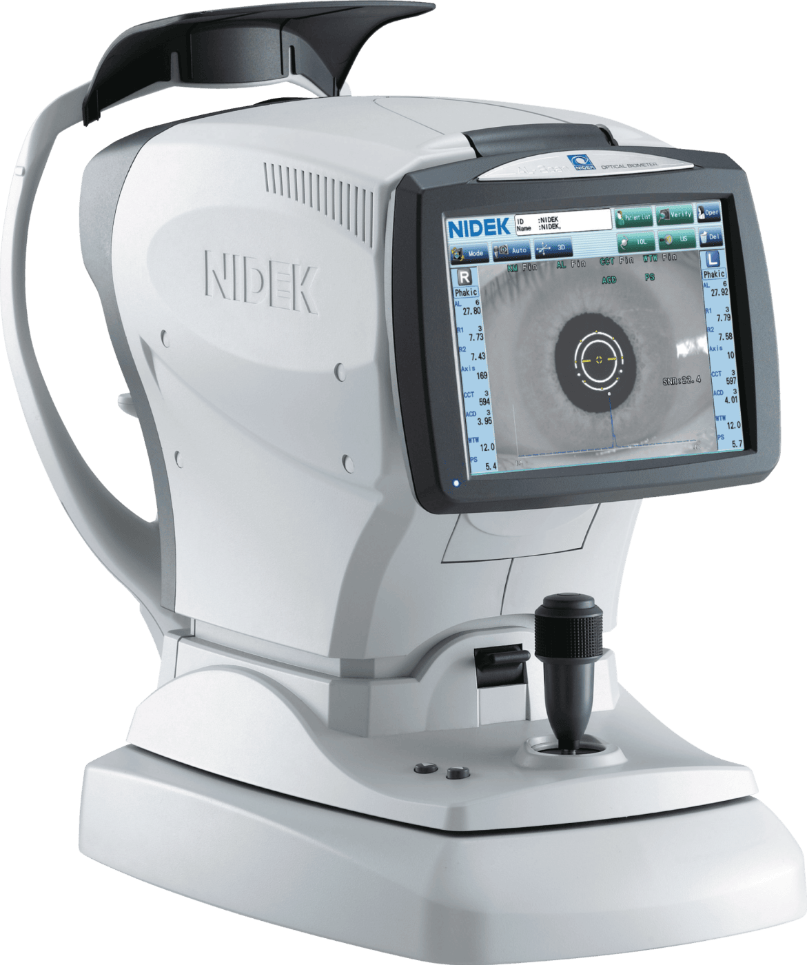
Built-In Touch Screen

Large Tiltable LCD screen for superior viewing angle and operational position options.

More details

Built-In Printer

The built-in printer provides an instant printout of the analyzed data and images of the endothelial cells.
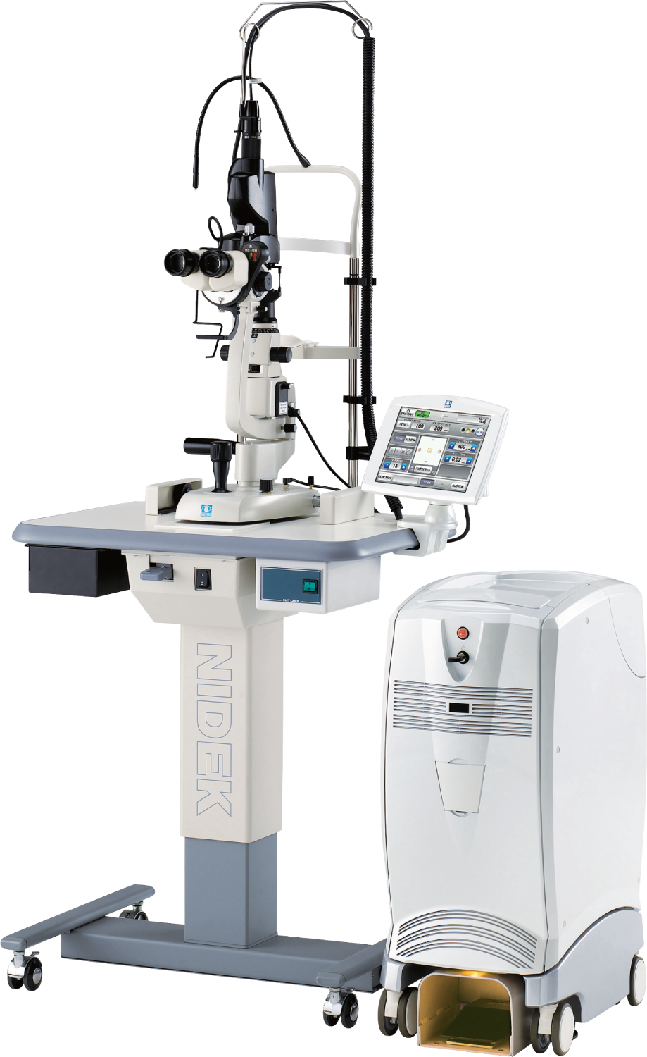
CEM-530 Overview

Gold Standard Technology

Unique Built-in Function: Paracentral Mode

The combination of central, paracentral, and peripheral imaging provides a broader, overall view that can be used for detailed morphological and quantitative evaluation of the endothelial layer and individual cells.

Seamless Imaging from Start to Finish

With 3D auto tracking and auto shot, vision practitioners can now easily choose optimal images for analyzing data, get comprehensive analysis in 2 seconds, and view endothelial cells in 4 different modes.

Enhance CEM-530 with CEM Viewer for NAVIS-EX

An optional function vision practitioners can choose is the CEM Viewer – a software for viewing and working with data using NAVIS-EX. Features include data management and endothelial cell count, progression follow-up and comparison, and paracentral display with peripheral.