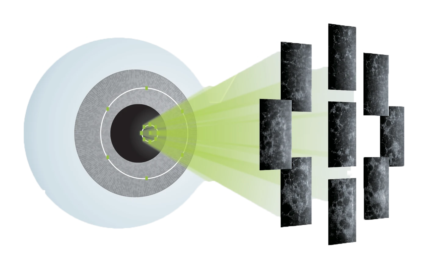
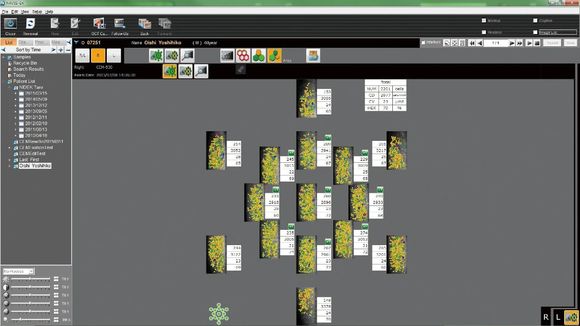
Paracentral Mode
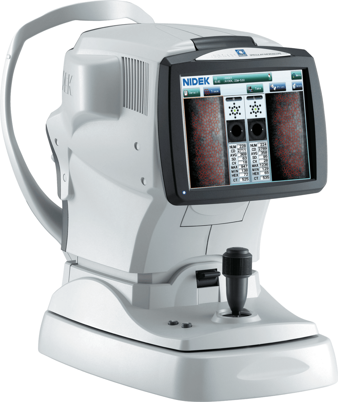
Multi-Area Specular Microscopy
The combination of central, paracentral (8 points), and peripheral (6 points) imaging provides a broader, overall view that can be used for detailed morphological and quantitative evaluation of the endothelial layer and individual cells.
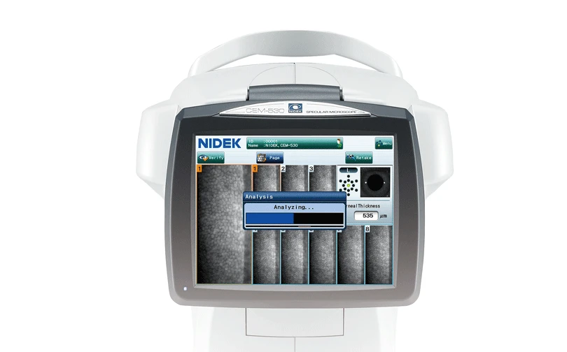
Two Second Auto Analysis
Seamless Auto & Manual Integrations
Rapid analysis increases the efficiency of the practice. Once the image is selected, complete analysis is automatically performed in two seconds with the CEM-530.
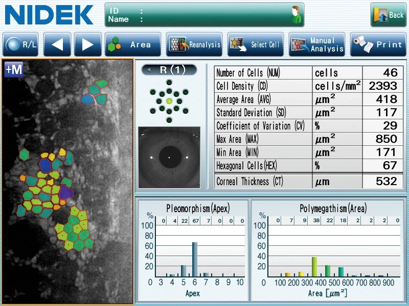
The analysis screen allows visualization of the endothelial cells in four modes, trace, photo, area, and apex, which helps the clinician to verify analysis values with the correspondent cell images.
2 Seconds
Auto Analysis with 4 Modes for Viewing Endothelial Cells
2 Histograms
In Shape (Pleomorphism) and Size (Polymegathism)¹
The CEM-530 provides comprehensive analysis including two histograms of variation in shape (pleomorphism) and size (polymegathism). For detailed analysis, the range of analysis can be changed and the cells to be excluded can be selected at the user’s discretion.
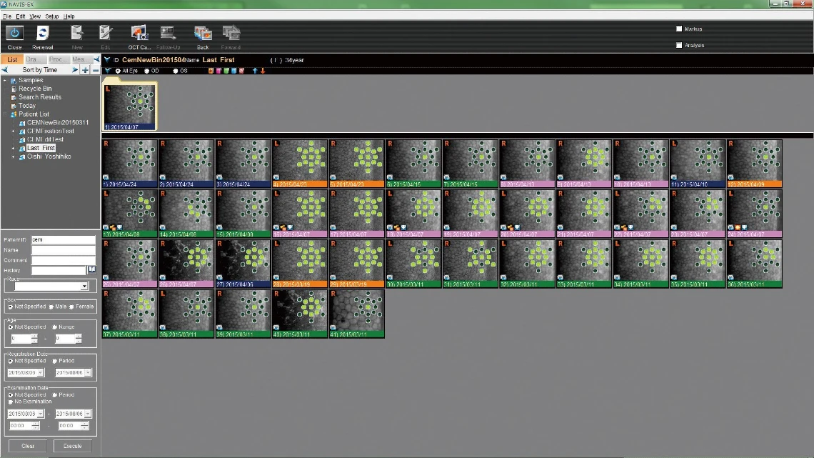
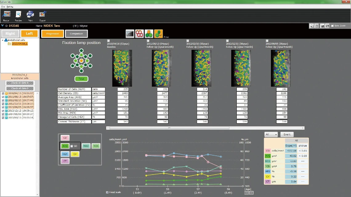
Optional
Get Additional Features with CEM Viewer for NAVIS-EX¹
CEM Viewer is software used for viewing and working with CEM-530 data via NAVIS-EX. This function enhances the capability of the CEM-530 with additional features and increases the efficiency of any clinic.
CEM-530
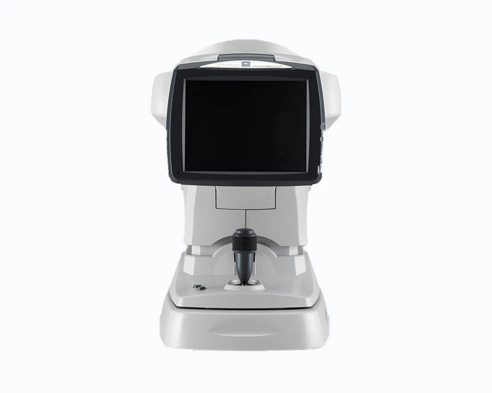
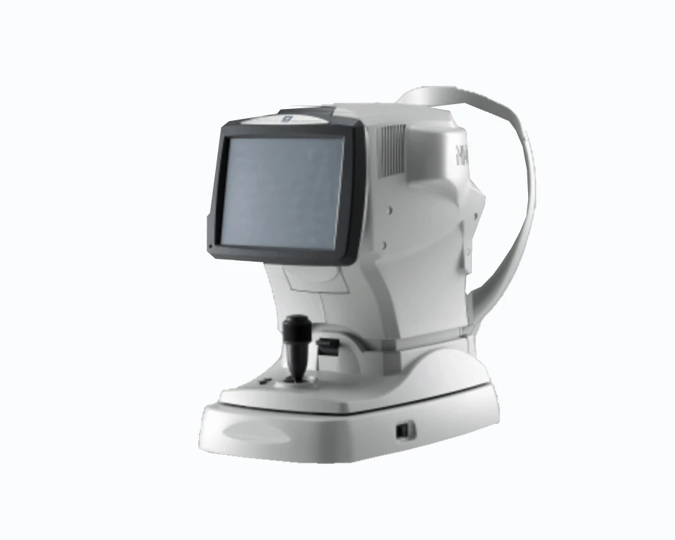
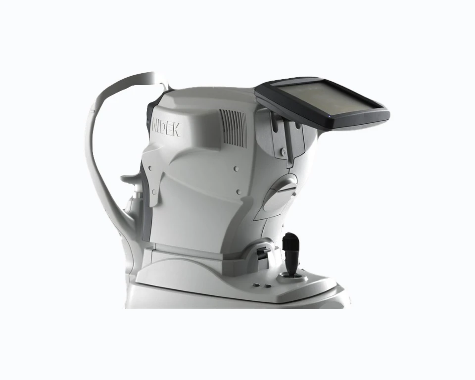
Experience a powerful specular microscopy and get access to premium support and resources.
Get in touch with a sales professional.
¹ NAVIS-EX is optional software and is not required for use of the CEM Viewer.