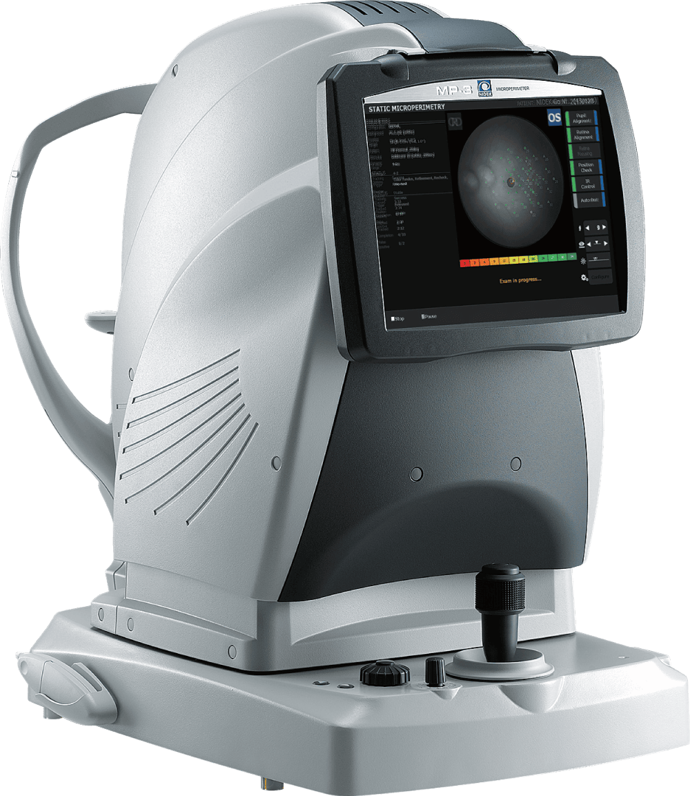
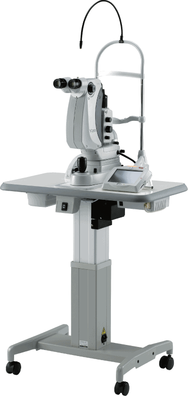
New Paradigm in Low Vision

The functional evaluation of retinal pathology is further advanced with the use of the microperimetry.

The functional evaluation of retinal pathology is further advanced with the use of the microperimetry.

Specifications

Device
Microperimeter

Model
MP-3 & MP-3 type S

Type
Diagnostic

Specialty
Retina & Glaucoma

Microperimetry

Visual field
40°

Maximum Stimulus Luminance
10,000 asb (in Accordance with ISO12866 Measurement Methods)

Background Luminance
31.4 asb / 4 asb (in Accordance with ISO12866 Measurement Methods)

Stimulus Size
Goldman I/II/III/IV/V Compatible

Threshold Strategy
4-2 / 4-2-1

Fixation Target
Shape: Single-Cross, Circle, Four-Crosses
Color: Select From White/Yellow/Red/Blue

Fundus Camera

Type
Non-Mydriatic Fundus Camera, Color

Angle of View
45° ±5% (The Refraction of the Eye is 0 D)

Diopter Correction Range
-25 to +15 D

Fundus Auto Focus Range
-12 to +15 D

Minimum Pupil Diameter
ø4.0 mm

Auto tracking
X-Y-Z direction

Auto shot
Available

Working Distance
45.7 mm

Display
10.4-inch color LCD touch screen

Camera
Built-in 12-megapixel CCD camera

Power
Supply: AC 100 to 240 V, 50/60 Hz
Consumption: 160 VA

Dimensions
Width: 13.1 in (334mm)
Depth: 22.1 in (562mm)
Height: 22.0 in (560mm)

Weight
79 lbs. (36kg)

Standard Add-Ons
NAVIS-EX software

What's Included
Limited Service Warranty
Complimentary Support
On-Site & Remote Assistance
Access to Learning Resources
start here

Support and Services
from World-Class Experts

Learn how you can prolong the lifetime of your investments while saving money and reducing costs.

* Caution: U.S. Federal Law restricts this device to sale, distribution, and use by or on the order of a physician or other licensed eye care practitioner.