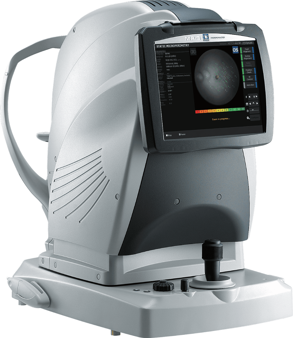
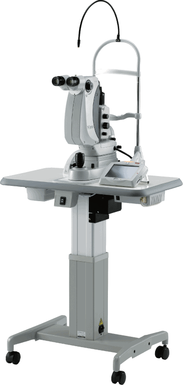
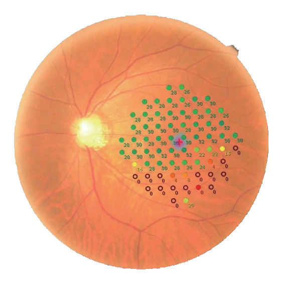
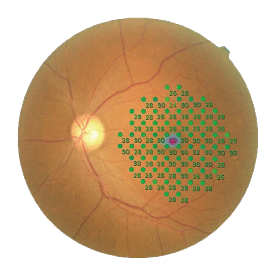
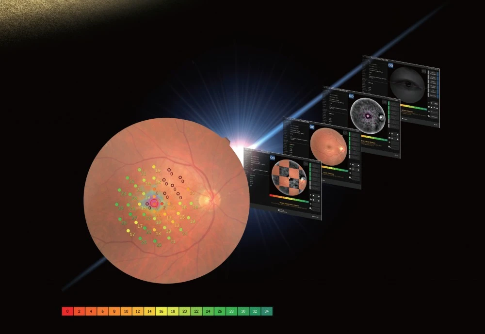
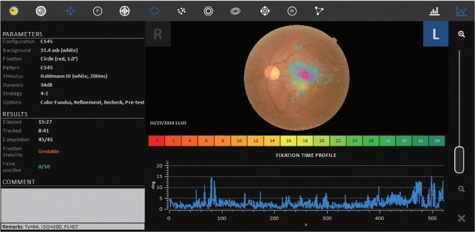
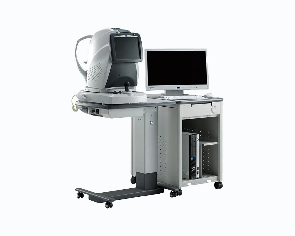
High Definition Measurements
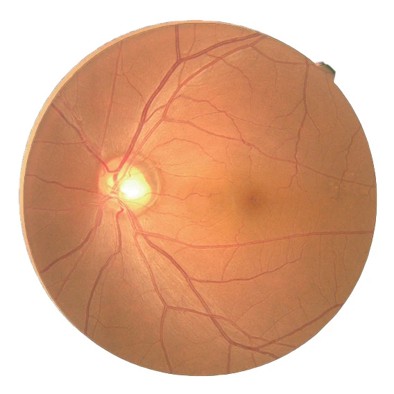
The MP-3 measures local retinal sensitivity for functional assessment of the retina. The results can be displayed over a color fundus image, correlating retinal anatomy to retinal function.
- Easy Image Acquisition with 12 Megapixel Camera
- Wider Range of Stimulus Intensity from 0 to 34 dB
- Perimetric Threshold Values, Even for Normal Eyes
Increase Comfort and Better Efficiency
Auto tracking and auto alignment functions provide more accurate measurements increasing patient and operator comfort and efficiency. These functions allow easy follow-up and reduce variations between examiners, resulting in well-aligned follow up exams.
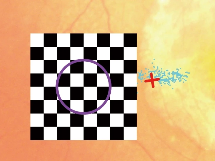
Feedback Exam for Visual Rehabilitation
The visual rehabilitation mode trains low-vision patients who have lost foveal fixation to relocate their preferred retinal locus (PRL) to a different region, called the trained retinal locus (TRL). The TRL is predetermined by a physician, and fixation rehabilitation allows the patient better functional vision (i.e. reading speed) due to increased fixation stability and visual outcomes.
Active flickering pattern stimulation and cheery music create an effective and pleasant training experience for the patient.
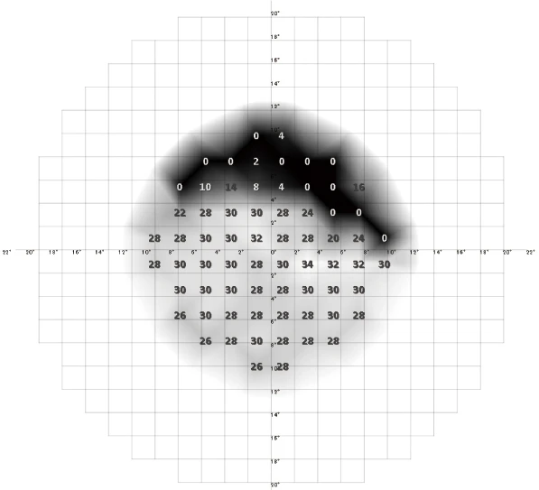
PRE- AND POST-TREATMENT COMPARISON
Vision Focused on Value for the Patient
Case of anti-VEGF treatment for age-related macular degeneration (AMD)
Pre-Treatment
Circle at 2° Percentage of Fixation Points 66.1%
Circle at 4° Percentage of Fixation Points 92.1%
Mean Sensitivity: 20.4
Post-Treatment
Circle at 2° Percentage of Fixation Points 68.1%
Circle at 4° Percentage of Fixation Points 95.5%
Mean Sensitivity: 20.9
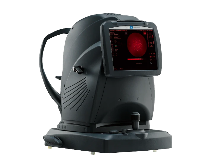
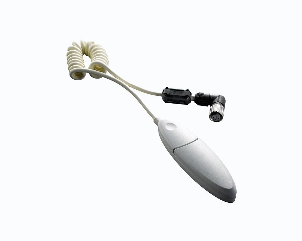
Additional features on the MP-3
FACTORY-INSTALLED OPTION
Scotopic Microperimetry
Scotopic microperimetry is used to assess the changes in rod sensitivity of degenerative retinal diseases including age-related macular degeneration and some forms of retinitis pigmentosa. The type S provides scotopic microperimetry in addition to the standard functions of the MP-3.
MP-3 &
MP-3 type S


Experience powerful digital technology and get access to premium support and resources.
Get in touch with a sales professional.