Skip to content

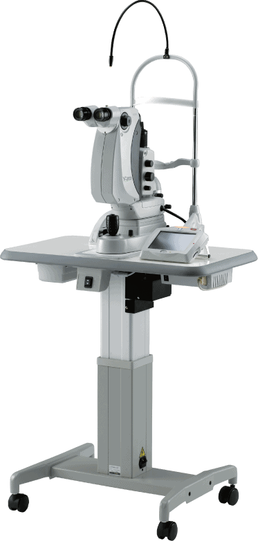
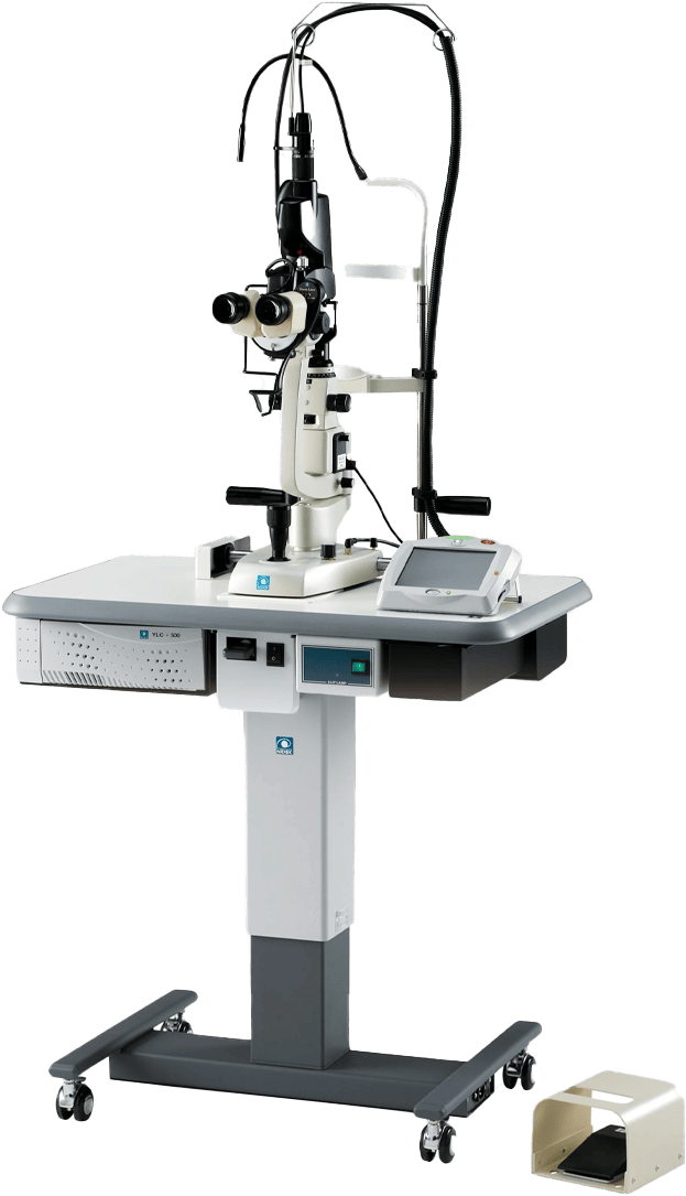
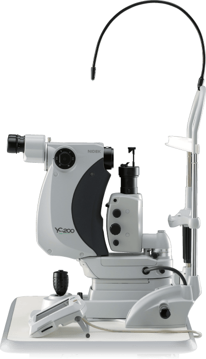
NIDEK Ophthalmic YAG & YAG/SLT Laser System YC-200
YC-200 S plus

Technology

Optional

Coaxial Illumination Tower

The YC-200/YC-200 S plus is capable of seeing and illuminating opacities in the vitreous with NEW optional coaxial illumination tower, which makes visual axis and illumination axis coaxial. The tower optimizes visualization during posterior membranectomy, capsulotomy and iridotomy and also illuminates vitreous opacities with the on axis/off axis illumination.

Optional

Combo YAG/SLT/Green

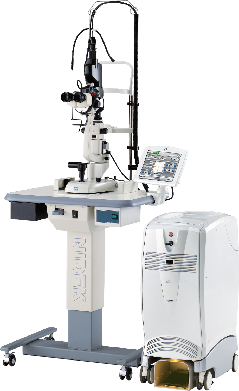
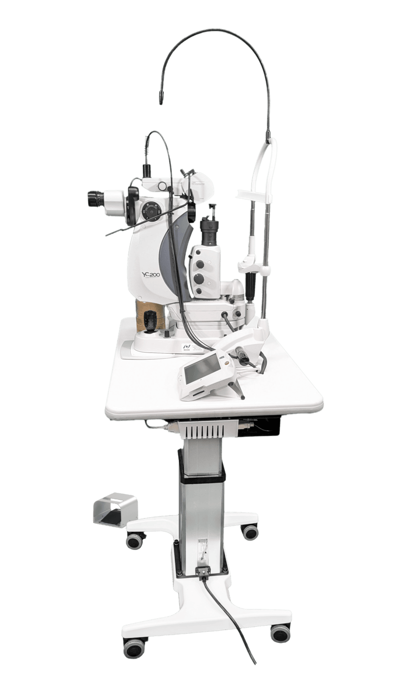
The YC-200/YC-200 S plus can be easily connected to NIDEK’s Green Laser Photocoagulator (GYC-500), allowing treatment of a wider range of patients and indications. Space requirements are minimized, and the NEW optional combination adapter includes an illumination tower (split mirror illumination).
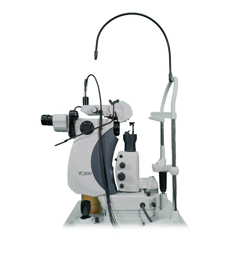
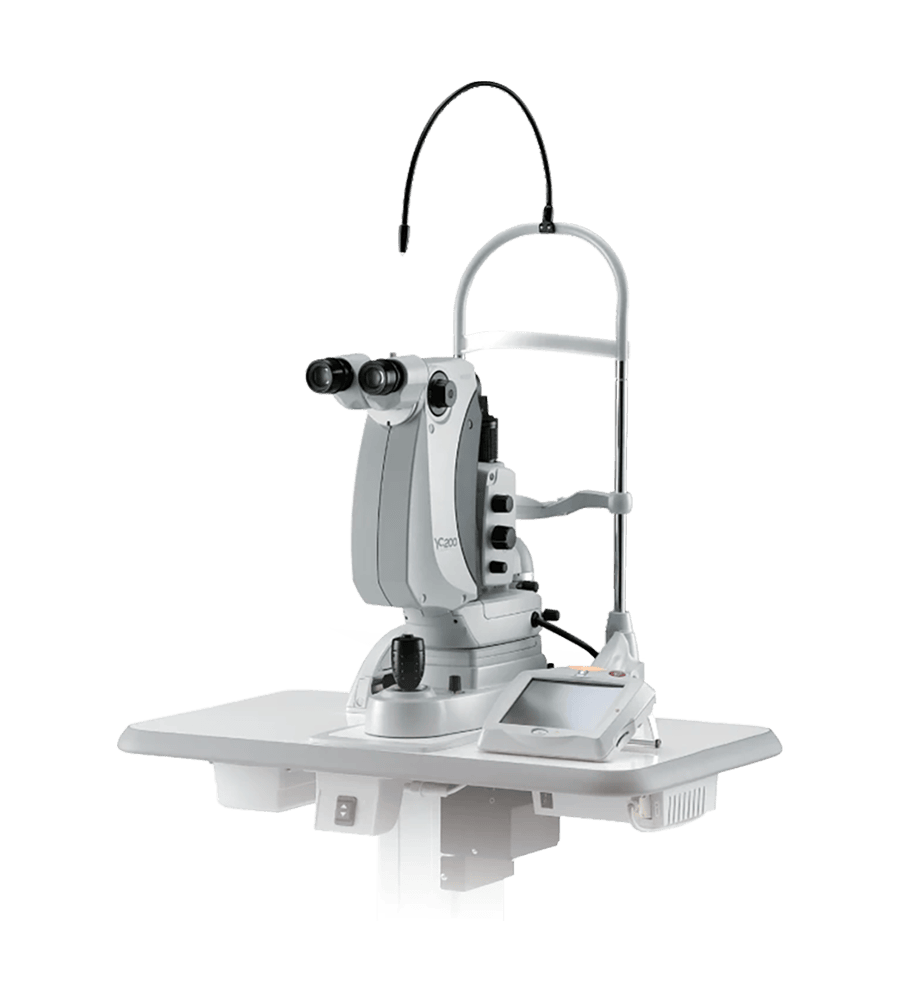
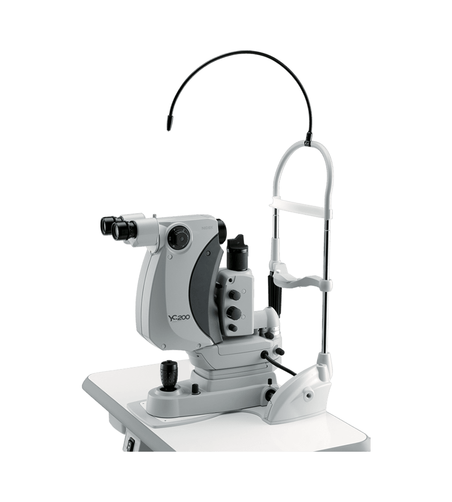
A Modern White And Gray Ophthalmic Laser Machine, Model Yc-200, Equipped With Various Adjustment Knobs, A Viewing Scope, And Flexible Cables, Designed For Eye Treatments. It Is Mounted On A White Base With Additional Attachments And Instruments Visible.

Refined Laser Delivery with Lower Energy

The YC-200 S plus / YC-200 achieves 1.6 mJ plasma threshold in air¹, delivering accurate and robust treatments with lower energy.

Precise Treatments

A suite of technologies has been incorporated in these lasers to achieve seamless function and greater precision. Features for targeting pathology, accurate energy delivery, and operative assist functions allow the surgeon to deliver treatments “Right on the Mark”.
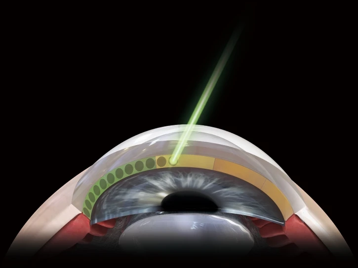
The YC-200 S plus offers an advanced SLT mode. SLT is highly effective for treating open angle glaucoma.

The SLT-NAVI feature presents an intuitive display on the progress of laser treatment, providing the surgeon with important feedback.

A Digital Illustration Of An Eye Undergoing A Nidek Laser Procedure. The Cross-Sectional View Shows A Laser Beam Targeting The Cornea, Creating Small Circular Incisions. The Background Is Black To Highlight The Surgical Process.
A Bar Chart Titled &Quot;Average Annual Energy Production Per Turbine&Quot; Shows Nidek'S 2010 Vs. 2040 Projections. For 2.5 Mw Turbines, 2010 Is 4,359, And 2040 Is 5,697. For 5 Mw Turbines, 2010 Is 5,128, And 2040 Is 10,749.

Cornea-friendly SLT

With a 5.5° cone angle, the YC-200 S plus is designed to decrease energy density on the cornea for sparing tissue from repeat treatments. The energy density on the cornea is reduced by more than one-third with a 5.5° cone angle compared to a 3.0° cone angle.
Exact Targeting

Clear and Sharp Field of View

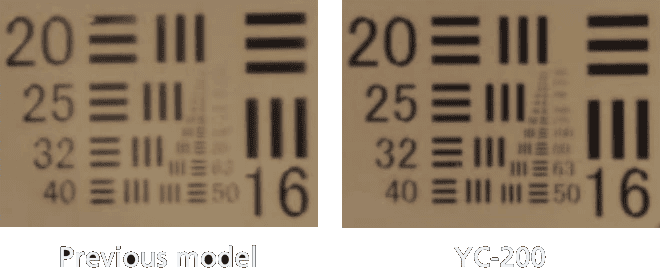
An improved optical design that optimizes resolution and contrast, and an expanded focal depth provide a clear view of pathology and treatment. A unique illumination system with an LED light source results in a bright, near-natural view that minimizes aberration. The sharp, clear optics provide an enhanced view of the treatment area.

Side-By-Side Comparison Showing Two Different Models: &Quot;Previous Model&Quot; On The Left And &Quot;Yc-200&Quot; By Nidek On The Right. Both Images Feature A Resolution Test Chart With Sets Of Horizontal And Vertical Lines In Various Patterns, Alongside Corresponding Numbers Indicating The Line Pairs Per Millimeter.
A Diagram Showing Two Blue Eyes, Each With Red Arrows Indicating Rotations. The Left Eye Has Arrows Pointing From The Top-Right To The Bottom-Left, While The Right Eye Has Arrows Pointing From The Bottom-Right To The Top-Left. A Circular Nidek Symbol Is In Between The Eyes.

Motorized Rotatable Aiming Beam

YAG Mode

The dual aiming beam offers superior targeting with a 360° rotating aiming beam that can avoid corneal opacities and achieve more accurate focusing.

Precise Edge Aiming Beam

SLT Mode

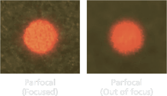
Parfocality delivers a clear view for easier focusing of the aiming beam through the contact lens.

Two Microscope Images Of A Red Circular Object Are Shown Side By Side With The Left Labeled &Quot;Parfocal (Focused)&Quot; And The Right Labeled &Quot;Parfocal (Out Of Focus).&Quot; The Left Image, Captured Using Nidek Equipment, Is Sharp And Clear, While The Right Image Is Blurry.
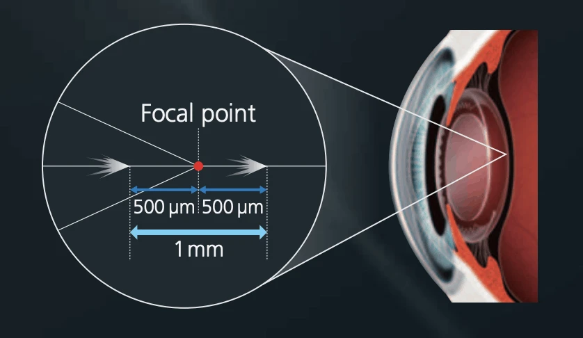
An Illustration Showing A Detailed View Of A Focal Point Within An Optical Element, With Dimensions Marked In Micrometers (500 Μm X 500 Μm) And Millimeters (1 Mm). The Optical Element, From Nidek, Is Magnified On The Right Side Of The Image, Highlighting Its Structure.

Wide Range of Focus Shift

The focus shift enables a 500 µm anterior or posterior axial shift of the focal point of the YAG laser. A change in 25 µm increments achieves precise treatments irrespective of the severity of pathology.

Smooth Operation Workflow
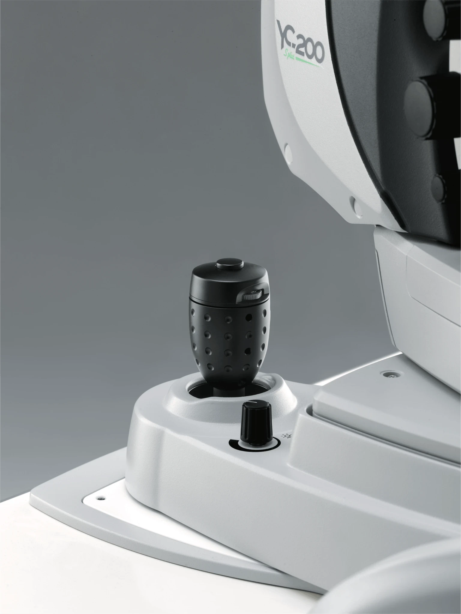
A Close-Up View Of A Grey Nidek Medical Device, Featuring A Joystick With A Dimpled Surface And A Small Black Control Knob On Its Base. The Background Is A Smooth, Gradient Grey, Emphasizing The Device'S Ergonomic Design And Precise Controls.

UNIQUE JOYSTICK

Utility S-switch

The S-switch on the joystick changes treatment settings without shifting gaze from the oculars. The ease of use allows surgeons an increased level of comfort during treatment.
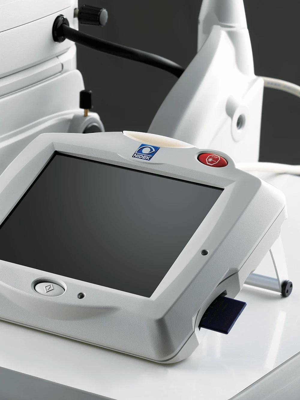
A Close-Up Of A Gray Nidek Medical Device With A Large Screen And Various Buttons, Including Prominent Green And Red Buttons On The Top. The Device Is Positioned On A White Surface, With Some Of Its Accompanying Equipment Partially Visible In The Background.

KEY CARD FUNCTIONALITY

Control Box

An intuitive graphic user interface and easy-to-read touch screen color LCD allow quick and easy setup and verification of treatment parameters. An SD card starts the unit, enables software upgrades, and saves treatment summaries.
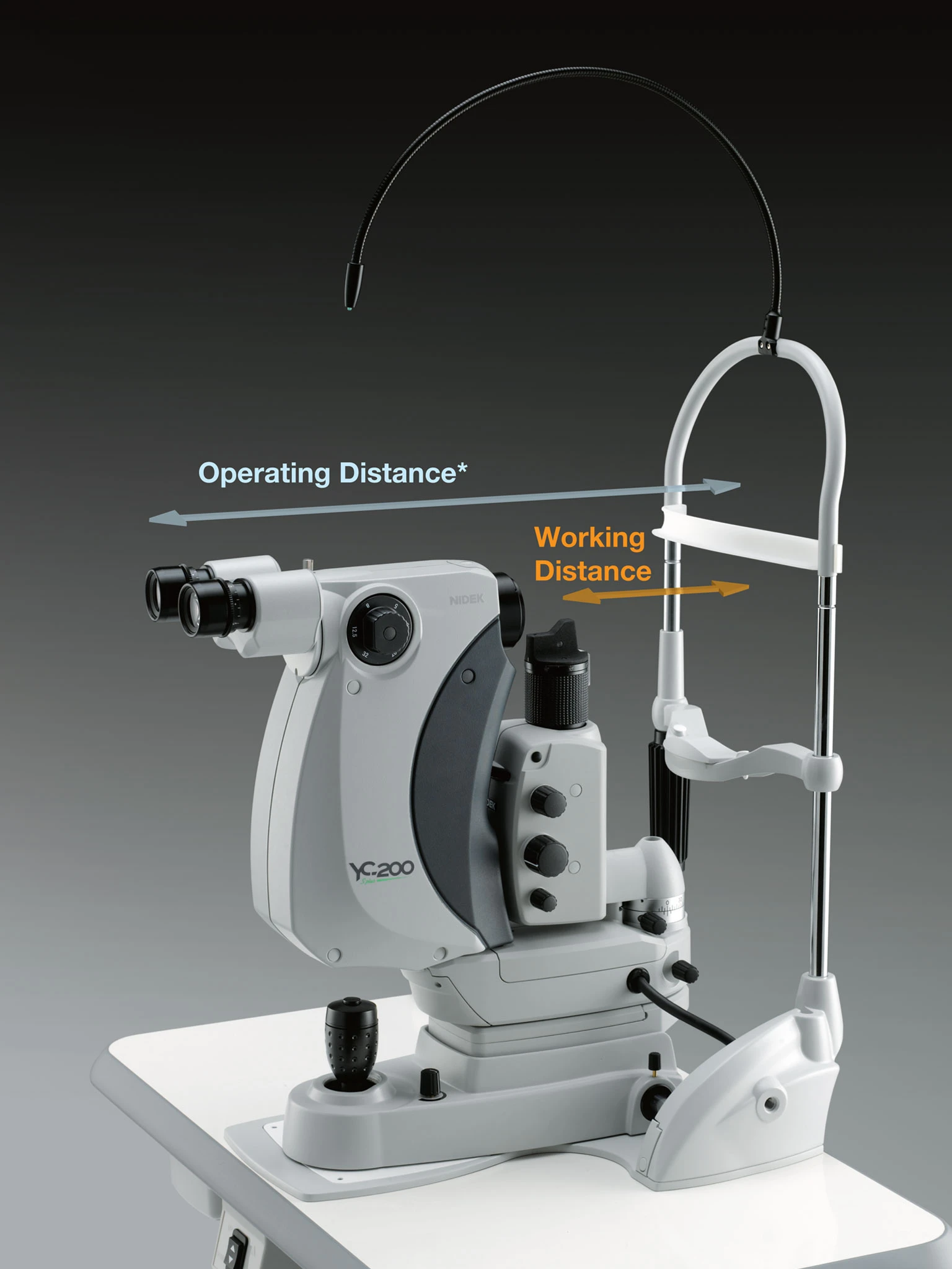
A Medical Device Used For Eye Examinations, Featuring Binocular Lenses And Various Controls. The Image Has Labeled Markers Indicating &Quot;Operating Distance&Quot; And &Quot;Working Distance.&Quot; The Nidek-Labeled Device, Model Xc-200, Is Mounted On A White Surface Against A Dark Background.

OPTIMIZED OPERATING DISTANCE

Slit Lamp

Optimized working distance allows easier manipulation of the contact lens, and the short operating distance decreases surgeon fatigue during treatment.

*Operating distance denotes the distance from the microscope eyepiece to the patient’s eye.

Experience powerful digital technology and get access to premium support and resources.

Get in touch with a sales professional.

¹ A plasma threshold of 1.6 mJ is achieved in ordinary room conditions (in-house data).
² The same laser delivery parameters were used on both samples of photo paper.
³ Data from theoretical simulations.
⁴ SLT Mode available for YC-200 S plus.