Your Vision is Our Mission
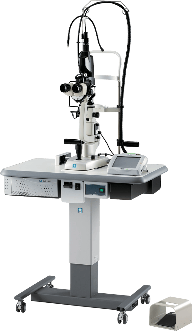
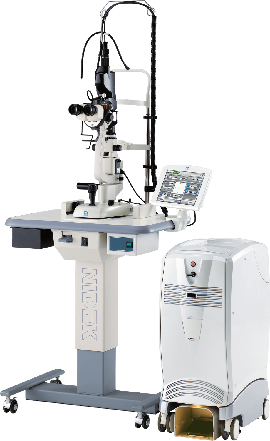
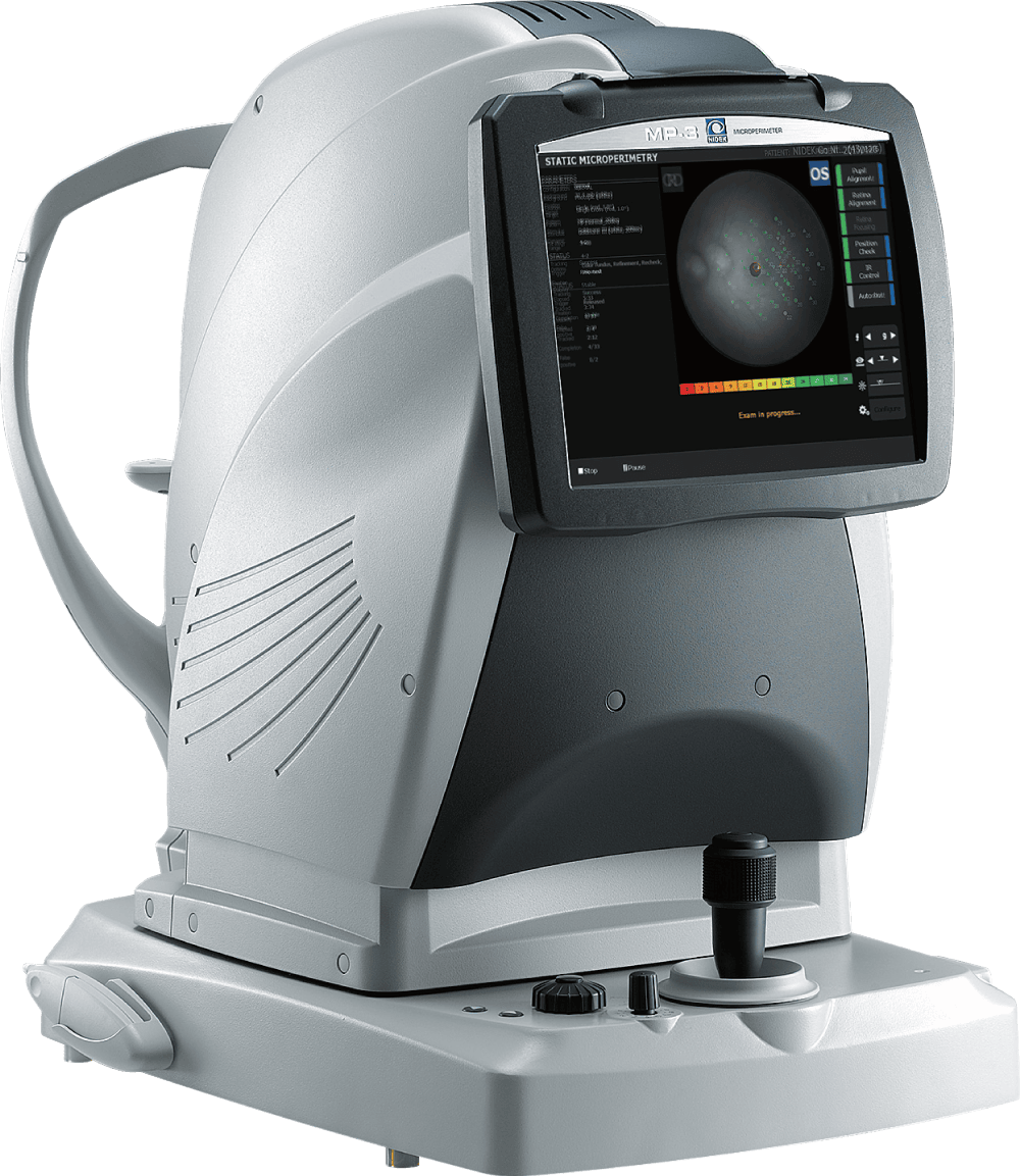
With a long history of research and development in examining, diagnosing, and treatment of eye disease, NIDEK deliver solutions to your needs at all stages of eye care.
Expanding the Boundaries of Vision Science
We have an unparalleled devotion to expanding the boundaries of vision science and contribute directly to shaping the future of vision technology, including the daily lives of patients and vision practitioners, as reflected by our ongoing research and development of new technologies optoelectronics and artificial vision.
Unrivaled "Made by NIDEK" Quality Assurance
NIDEK is proud that our quality assurance activities gained the international recognition that these international certifications attest. We will continue to strive to enhance our product quality even further and deliver products that our customers can use with confidence.
Do you recognize this balloon?

If you have seen this balloon in an optical device, then you have used a NIDEK product! You were able to experience an examination by the leading technology used for measuring the refraction of eyes, which can determine reference values for how much corrective power you will need in your glasses or contact lens.
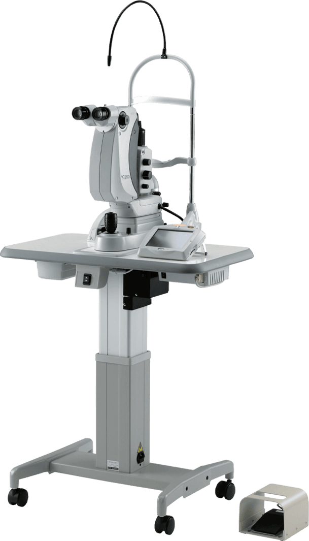
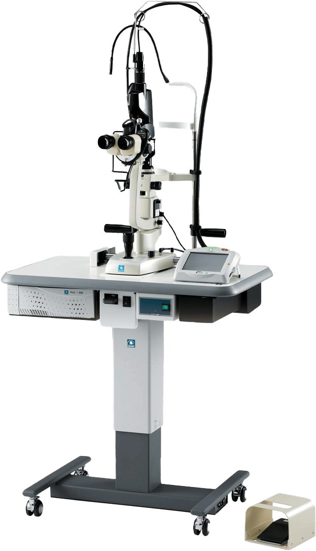
NIDEK is changing how we see
Preparing Vision Practitioners with Innovations for Today and Tomorrow.
We help eye care professionals around the world not just with better eye care technology, but with a better eye care experience. Our technological advancements provide reliable ophthalmic equipment that focuses on value for doctors and their patients.
AS FEATURED IN