 More details
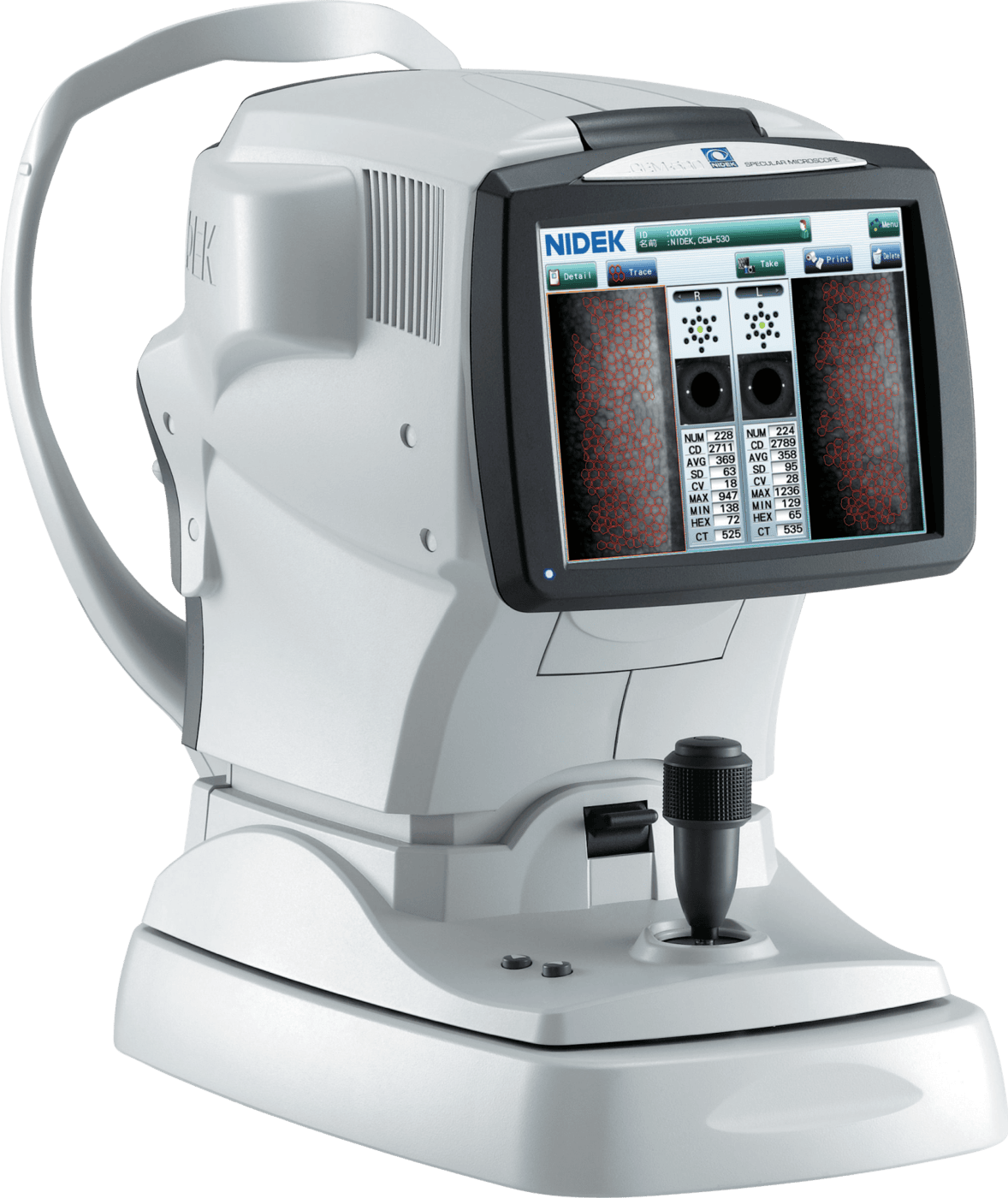
Multi Area Specular Microscopy
The combination of central, paracentral, and peripheral imaging is available.
Patient Comfort
The 3-D auto tracking and auto shot, provides ease of use, allowing faster and more accurate measurement.
Built-In Touch Screen
Large Tiltable LCD screen for superior viewing angle and operational position options.
Built-In Printer
The built-in printer provides an instant printout of the analyzed data and images of the endothelial cells.
CEM-530 Overview
Gold Standard Technology
Get Started
Take Patient Care to the Next Level
Accelerate the discovery and development of patient treatment and operate more efficiently with NIDEK’s ophthalmic solutions.
